An aural hematoma is a collection of blood within the cartilage of the ear and the skin. This is most commonly the result of external physical trauma to the pinna (the “flap” of the ear) or self-inflicted injury from your pet’s scratching and head shaking secondary to infection of the external ear canal. The condition is common in dogs with chronic otitis externa and less common in cats. Hematoma formation has also been associated with increased capillary fragility (e.g., as seen with Cushing’s disease).
Sources of irritation to the ear linked to the development of an aural hematoma include:
- inflammation
- immune mediated diseases
- allergies
- parasites
- foreign bodies
- trauma (bite wound or blunt trauma)
Most animals usually have an associated infection. Recurrence of the condition is common if the underlying condition is not resolved.
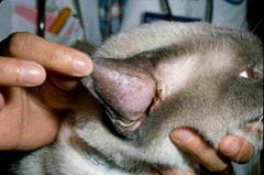
Swelling associated with an aural hematoma is most apparent on the concave inner surface of the pinna (Figure 1). The swelling is soft, fluid-like and warm in the early stages. With time, scar tissue will thicken and deform the ear, resulting in a cauliflower contracture.
A few simple tests may be performed to make sure there is not an underlying reason for the ear being irritated or hard to heal from the bleed.
- fine needle aspirate and cytology
- systemic testing for underlying causes may include
- allergy testing
- ear swabs
- endocrine testing
Pet owners should contact an ACVS board-certified surgeon for advice early in the disease process before chronic changes occur in order to achieve the best results. Treatment options include needle aspiration and bandages, tube drainage systems, incisional drainage and/or tacking sutures to eliminate the space that fluid can accumulate. The goals of surgery are to remove the hematoma, prevent recurrence and retain the natural appearance of the ears. Surgery typically includes making an incision on the underside of the ear flap to drain the fluid and followed by placing several sutures to prevent fluid from building back up. A bandage is typically placed after surgery for days to weeks to help decrease swelling, discharge, and trauma.
Deformity of the ear can occur if the condition is left untreated. This typically will leave the animal with a “cauliflower” ear. Potential complications include:
- cosmetic alteration of the ear
- recurrence of the hematoma
- necrosis (death) of the pinna
A bandage should be placed to protect the ear from infection and self-inflicted trauma. Infection can occur in the surgical site if surgical wound is not managed appropriately with bandages.
Aural hematomas seldom recur if they are properly treated and the underlying disease is appropriately addressed. This condition can be prevented by providing prompt attention to conditions that result in irritation of the ears.
Premier Sponsors


