The umbilicus in newborn calves consists of the urachus, a tube that connects the fetal bladder to the placental sac, and the remnants of the umbilical vessels that transported blood between the fetus and its mother. Normally, just after birth, these structures shrink until only tiny remnants remain within the abdomen. If bacteria gain entry through the umbilicus, however, those remnants can become infected and require surgical removal. Additionally, if the area in the body wall through which these structures passed remains open, the intestines can protrude through the defect, resulting in an umbilical hernia. Umbilical hernias are the most common birth defect in calves and may be more common in the Holstein-Friesian breed.
Calves with simple hernias may seem completely normal except for a reducible umbilical hernia (where the hernia contents can be easily pushed back into the abdomen). Calves with infected hernias may be sick and show signs such as fever, inappetence, and poor growth rates.
If the umbilical vessel remnants are involved you may see frequent urination in small amounts and urination through the umbilicus. Infection of the umbilical vein can spread to the liver, causing fever, and poor growth rates.
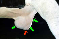
Calves are physically examined to try to reduce the hernia contents. In some instances, it is possible to palpate an infected stalk within the hernia sac or around the umbilicus (Figure 1). Sedation may be required to place the calf on their back or side to better feel these structures.
Ultrasound examination is useful in determining whether there is any infection present and how extensive it is.
Laboratory work such as white blood cell counts or fluid collection from the mass for a bacterial culture can also be useful detecting infection.
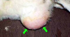
Routine hernias can be repaired in the field, but those with extensive infection are best repaired by an experienced ACVS board-certified veterinary surgeon at a referral hospital where general anesthesia is available. Surgical repair is the treatment of choice. Surgery can occasionally perform under sedation with local nerve blocks, but general anesthesia is preferred. The umbilicus and its associated structures and all infected tissue are removed. When the infection extends all the way to the bladder, a portion of the bladder wall may need to be removed. If a large abscess is present it may have to be drained before surgery can be done (Figure 2).
For simple hernia repairs antibiotics are often given only at the time of surgery. However, if there is an extensive infection, a longer course of antibiotics may be necessary. Non-steroidal anti-inflammatory drugs are useful for reducing pain but should be used cautiously since they can cause abomasal (stomach) ulcers if given too long. After surgery, the calf should be slowly re-introduced to feed and confined, to some extent, to prevent tension on the surgical repair.
Prognosis is favorable for recovery, especially for simple hernias or minor infections. Calves that have infected umbilical vein remnants extending towards the liver are at an increased risk of peritonitis (infection of the abdominal cavity) and therefore have a poorer prognosis.
Premier Sponsors


