Indications for ovariectomy in the mare include removal of benign tumors or to achieve behavioral modification related to estrus. Many surgical approaches and techniques have been described, including removal under general anesthesia in dorsal recumbency (positioned on the back), standing through a flank approach, or removal through the vagina with a crushing-type instrument. Laparoscopic (keyhole) removal of ovaries has become a popular alternative as it is minimally invasive, avoids the risk of general anesthesia, greatly improves visualization and access to the base of the ovary and its blood supply (pedicle), and provides a cosmetically superior result with decreased convalescence.
The most common reason for ovariectomy is removal of benign tumors affecting the ovary called granulosa thecal cell tumor (GTCT). These tumors are discovered because of aggressive behavior, including stallion–like tendencies, toward other horses and people.
Additionally, ovary removal can attenuate persistent signs of estrus (being in season) when other forms of medical and hormonal therapies have been attempted. There is some controversy and difference in opinion regarding the success of this procedure to provide modification in behavior.
Diagnosis is based on recognition of aggressive clinical signs, ovarian palpation and ultrasound and confirmation with blood work evaluating elevated levels of anti-mullerian hormone, testosterone and inhibin circulating in the bloodstream.
Standing laparoscopic ovariectomy allows for direct visualization and access to the target organ. The procedure provides a cosmetic result with a reduced period of convalescence.
Pre-Operative Preparation:
Since the ovaries are located in the abdomen, immediately adjacent to the gastrointestinal tract, it is important to withhold feed for at least 24–48 hours to allow for visualization of the ovaries, which lie suspended toward the back of the horse near the pelvis.
Laparoscopic Ovariectomy Procedure:
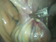
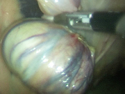
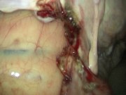
The mare is sedated and restrained in stocks. The hair on the flank on the affected side is clipped, sterilely prepared, draped and blocked with local anesthetic. Several small (1.5 cm) incisions are made within the flank region and specialized trocars or rigid tubes are placed into the abdomen allowing introduction of the laparoscope (rigid long camera) into the abdomen, so that internal structures can be visualized (Figure 1). Instruments and devices to grasp and cut the ovary while sealing the blood vessels are placed through the remaining portals. Occasionally, the abdomen needs to be insufflated or expanded with carbon dioxide (CO2) gas to allow for better visualization of the ovary and its attachments to the uterus. The base of the ovary is locally anesthetized, which is followed by surgical removal (Figure 2). The surgical site is then observed to verify no persistent bleeding is present (Figure 3). Occasionally, the ovary is too large to remove and may need to be transected and removed in stages. Additionally, if the ovary is too large, a standard ventral midline approach under general anesthesia may be elected
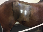
Postoperatively, the mare is maintained on antibiotics and anti-inflammatories for 2–5 days. Occasionally laparoscopy can cause an inflammatory response resulting in low-grade fevers. This is usually short-lived and resolves within 48 hours. Food is gradually reintroduced over the next few days. Incisions heal within two weeks and horses return to work typically within one month (Figure 4).
Premier Sponsors


