Rib fractures can occur more commonly than expected following a “normal” foaling or dystocia. The severity of which can vary from unknown to life-threatening depending on the amount of displacement of the fracture ends and location of the fracture i.e. over the heart. The size of the foal’s chest in relation to the mare would be a logical cause for rib fractures during foaling, however this has not been proven. It seems to occur due to overzealous attempts to deliver the foal before it is properly positioned. Perhaps the most apparent reason is incomplete extension of the elbows before the foal engages the birth canal. The reasoning is two-fold: elbow flexion places the elbow joint at the most common location for rib fracture (the 3rd-6th ribs) and increases the pressure on the rib cage at that location as the foal enters the birth canal potentially leading to rib fracture.
Foals have varying signs that depend on the severity of the rib fracture(s) including the number of ribs effected, location, and degree of displacement. Affected foals may have:
- Difficulty breathing
- Increase breathing rate
- Difficulty to stand
- Signs of abdominal discomfort or colic
- Lameness
- Swelling associated with fracture
- Physical examination with careful attention to palpation of each rib feeling for a step formation, break in the rib contour, or clicking of fracture ends
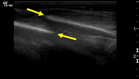
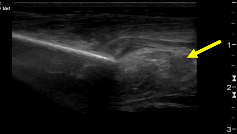
- Chest ultrasound can be the most useful method to assess the ribs, lungs, heart, and chest cavity to assess for fluid in the chest or surrounding the heart (Figure 1a and 1b)
- Radiographs of the chest
- Bloodwork
Conservative Management
One or two mildly displaced rib fractures that do not cause clinical signs such as increased respiratory effort can be managed conservatively. Conservative management entails stall or stall-sized pen rest for 3-4 weeks until a callus is palpable suggesting the fracture is stabilized enough to allow for small paddock turnout.
Surgical Management
Surgical management may be indicated in rib fractures that are markedly displaced, located over the heart, or there is damage to the lungs, heart, or diaphragm. Ultrasound findings in those cases may include increased fluid or blood in the chest cavity or in the sac surrounding the heart as well as intestines in the chest cavity if the diaphragm, which is a primarily muscular tissue that divides the abdominal and thoracic cavity, is perforated by a rib fracture.
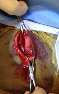
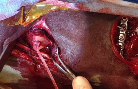
Surgical management is aimed to stabilize the rib fracture to prevent further displacement and reduce further damage to vital organs (Figure 2). Patients may be critically ill and should be stabilized prior to general anesthesia which may include a blood transfusion, intravenous fluids, supplemental oxygen, or other treatments if they have concurrent issues aside from fractured ribs.
Surgical Repair Techniques
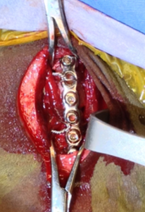
- Plate and screws fixed to the rib and further secured with a loop of cerclage wire around the rib to hold the plate in place (Figure 3)
- Nylon cable tie passed through a hole drilled through each fractured end and zipped closed to stabilize the fracture (Figure 4)
- Cerclage wire in a figure 8 pattern through the fracture ends to stabilize the fracture
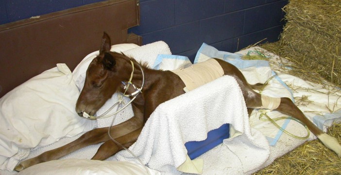
Foals with rib fractures that are mildly displaced and able to be managed conservatively can generally do very well. Surgically managed foals can also do well as long as there is no major secondary damage to vital organs. Post-operatively, they may require hospitalization in an intensive care unit until they are healthy enough to be discharged (Figure 5). Unpublished data showed that around 80% of both conservatively and surgically managed foals hospitalized in an intensive care unit survived to discharged. Surgical implants are typically left in place unless they become infected and these foals can have similar athletic performance compared to their siblings.
Premier Sponsors


