Granulosa cell tumors (GCT) are the most common type of tumor to affect the equine reproductive tract, most specifically the ovaries. These tumors are often benign (do not spread), however they can be performance-limiting due to associated behavioral issues and potentially pain depending on their size. These tumors can also affect the reproductive capability of a mare intended for breeding due to elevations in reproductive hormones. Typically, only one ovary is affected with a granulosa cell tumor, and the other ovary will be found to be smaller in size and inactive due to hormones that are being produced by the affected ovary. Treatment by surgical removal is curative, and following treatment, the unaffected ovary should be able to serve as a fully functional reproductive organ.
Common clinical signs associated with granulosa cell tumors include:
- Persistent and unexpected behavioral changes such as stallion-like behavior and aggression
- Intermittent or prolonged heat cycles (continuous estrus)
- No heat cycles (anestrus)
- Difficulty getting bred
These changes are due to an increase in the excretion of specific reproductive hormones by the tumor, such as testosterone and inhibin. Granulosa cell tumors can be found in mares of any age, but the average age of affected mares is 11 years.
Rarely, bleeding from a granulosa cell tumor can result in clinical signs consistent with hemoabdomen (blood in the abdomen). These signs may include but are not limited to: pale mucous membranes, quiet demeanor, increased heart rate, and colic. If you notice any of these clinical signs, please contact your primary care veterinarian immediately.
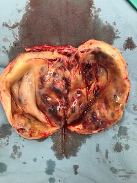
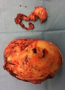
Granulosa cell tumors may be suspected based on behavioral changes and if your veterinarian finds an enlarged ovary on transrectal palpation. Additionally, the other ovary may be smaller in size than the affected ovary (Figure 1). Additional diagnostics such as transrectal ultrasound and hormone level testing are required to obtain a definitive diagnosis.
Often, ultrasonographic appearance includes enlargement of the ovary with a cystic or “honey-combed” appearance (Figure 2), however the appearance can be variable. Determination of reproductive hormone levels, specifically Anti-Mullerian hormone, inhibin, and testosterone, from a blood sample is recommended for definitive diagnosis.
Ovariectomy, also known as surgical removal of the affected ovary, is the treatment of choice to resolve this condition. In some horses, it may be recommended that both ovaries are removed during the same surgery. There are a number of surgical approaches that can be used for removal of the ovary and consultation with a board-certified veterinary surgeon will help determine which approach is best for your horse.
Most commonly, ovariectomy is performed laparoscopically (using a camera and specialized equipment) through a flank approach with the horse standing and sedated. This approach will require your mare to be held off feed for a period of time prior to the procedure in order to decrease the fill of her gastrointestinal tract. This approach is advantageous because it allows the surgeon to have better access to the ovary and blood supply for removal and avoids the need for general anesthesia. If an ovary is too large to be removed through a flank incision, your board-certified veterinary surgeon may recommend a two-part procedure where a portion of the surgery is performed laparoscopically and the ovary is removed through a ventral midline incision with the horse under general anesthesia.
Initial care after surgery depends on the size of the ovary and size of the incisions required to remove the ovary. Specific after care instructions will be made by your board-certified veterinary surgeon.
Generally, after care includes a period of stall rest with hand walking (10-14 days) with a gradual increase in exercise and turnout following this time period. Medications, such as antibiotics and anti-inflammatories, may also be recommended during the initial post-operative period.
As with any surgical procedure, there is always a potential for complications. With this type of procedure, complications may include:
- Intra-operative or postoperative hemorrhage from the area where the ovary was removed
- Inadvertant damage to the gastrointestinal tract when inserting laparoscopic equipment
- Post operative colic
- Swelling at the surgical site
- Infection of the surgical site
- Dehiscence, or opening, of the surgical incision
With the removal of one affected ovary, the normal ovary should resume normal reproductive function; however, this may take up to 6-8 months, depending on your mare and the time of the year. Ultimately, a large majority of mares are able to return to normal reproductive function, can become pregnant, and can carry a pregnancy to full term with the remaining ovary.
Premier Sponsors


