Cryptorchidism means, “hidden testes” (crypt = hidden, orchid = testes).

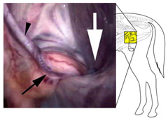
This term describes the condition in which one (unilateral) or both (bilateral) testicles do not descend normally into the scrotum. Generally, unilateral cryptorchids are usually fertile, while bilateral cryptorchids are generally sterile. The retained testicle may be located anywhere from within the abdomen to within the inguinal canal, which is the normal passage route into the scrotum (Figure 1). A single cause of equine cryptorchidism has not been established and contributing causes remain obscure. The condition is likely the result of a complex combination of genetic, hormonal, and mechanical factors.
The prevalence of left and right testicular retention is nearly equal, though retained left testes are more often in the abdomen, while the right retained testicle is more often in the inguinal canal. All breeds of horses may exhibit cryptorchidism, but there is a higher frequency in Quarter Horses, Saddlebreds, Percherons, and ponies. The condition is considered heritable, so affected pets should be gelded to help prevent the continuation of this congenital defect and for safety/behavioral reasons (although a testicle is undescended, it still produces male hormones leading to characteristic stallion behavior). Also, many breed associations do not allow registration of cryptorchids.
Cryptorchid horses usually exhibit standard stallion behavior, but visibly/palpably lack one or both scrotal testicles.
- Immature horses may be undetected until they are examined just prior to routine castration.
- Mature horses with no detectable testes that behave like stallions may be a:
- Bilateral cryptorchid
- Unilateral cryptorchid with the descended testes previously removed
- Geldings with stallion-like behavior (castrated later in life +/- previous breeding stallion).
Monorchidism (complete absence of one testicle) is rare in the horse and should only be considered after extensive testing and, potentially, surgical exploration.
Your primary care veterinarian may recommend performing the following diagnostics:
- Combination of external and rectal palpation +/- ultrasonographic examination to locate a testicle within the abdomen or inguinal canal.
- Blood tests measuring testosterone and conjugated estrogens when there is incomplete surgical history or absence of externally palpable testicles (~95% accurate, both tests should be performed if either is inconclusive).
- Testosterone levels: Measured in the blood before and after administration of human chorionic gonadotropin (hCG).
- Stallions and cryptorchids have higher levels of testosterone, and levels of the hormone increase after hCG administration.
- Castrated horses have low levels of testosterone that do not increase after hCG administration.
- Conjugated estrogen levels: Typically, levels are higher in horses with testicular tissue, and a single measurement can often identify cryptorchids.
- Unreliable in horses younger than 3 years and in donkeys.
- Testosterone levels: Measured in the blood before and after administration of human chorionic gonadotropin (hCG).
An ACVS board-certified veterinary surgeon should perform the identification and surgical removal of undescended testicles. The anatomy in this area is very complex, and testicles are often smaller than normal and may be abnormally formed/shaped with an abnormal appearance. Veterinary surgeons trained according to the standards of the American College of Veterinary Surgeons have specific knowledge and skills for the diagnosis and treatment of cryptorchidism in horses.
- Surgical removal of both testicles:
- Standard surgical approach: With the horse on their back and under general anesthesia, an incision is made, generally over the external inguinal ring. The testicular tissue is carefully located and manually removed in its entirety from the abdomen or inguinal canal. The external inguinal may be closed, and the incision closed routinely.
- Laparoscopic approach: With the horse under standing sedation or with the horse on their back under general anesthesia, using a camera and specialized equipment, the abdomen is distended with sterile carbon dioxide gas, and a camera is inserted into the abdomen through a small incision in the flank (standing) or umbilicus (on their back under general anesthesia). Once the testicle is located, additional small incisions are made to pass instruments into the abdomen for removal of the testicle.
With either approach, meticulous attention is paid to ensure that all testicular tissue is removed and the blood supply to the testicle is securely closed off prior to removal to prevent potential bleeding.
Initial care after surgery depends on the surgical procedure elected (open approach vs. laparoscopic approach), and specific discharge instructions will be given by your ACVS board-certified surgeon. The open approach usually consists of stall rest with hand walking for a short period of time with gradual increase in exercise after approximately 10 days to two weeks. Any external sutures are typically removed around 10–14 days after surgery.
Following laparoscopic surgery, the aftercare period is much reduced, and horses can resume turnout and light exercise after the first 72 hours, with external suture removal around 10–14 days.
As with any castration procedure, appropriate handling and socialization measures should be taken at the farm.
As with any surgical procedure, there are always risks of complications which may include:
- Complications while under general anesthesia
- Excessive/uncontrolled hemorrhage
- Inadvertent damage to the gastrointestinal
- Surgical site infection
- Post-operative swelling
- Incisional site breakdown, usually secondary to infection and swelling
Though hormone levels dissipate almost immediately after testicle removal, learned stallion behaviors often take a period of time and training to change and could persist indefinitely, depending on the horse.


