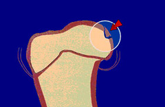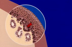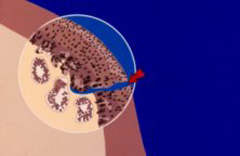
Osteochondrosis occurs commonly in the shoulders of immature, large, and giant-breed dogs. The lesion usually appears on the caudal (back) surface of the humeral head (Figure 1). Osteochondrosis begins with a failure of immature cartilage to form bone in the humeral head. This failure leads to abnormal cartilage thickening (Figure 2). Increased cartilage thickness may result in malnourished cartilage cells that die. Loss of these cartilage cells deep in the cartilage layers leads to formation of a defect at the junction between cartilage and bone. Subsequently, normal daily activity may cause fissures in the cartilage that eventually communicate with the joint, forming a cartilage flap (Figure 3). It is with the formation of a flap that osteochondrosis becomes osteochondritis dissecans (OCD). OCD is the form of osteochondrosis that is associated with pain and dysfunction. In some cases, the resulting flap occupies as much as half the humeral head. The cartilage flap may completely detach from the underlying bone and become lodged in the back of the joint pouch. Free cartilage flaps can lodge in joints and may increase in size with calcification becoming “joint mice” which can be seen on radiographs.

The causes of OCD are multifactorial with genetic and nutritional interactions thought to be the central factors. Risk factors for OCD may include:
- Breed genetics (Polygenic trait)
- Age
- Gender
- Anatomic abnormalities
- Rapid growth
- Nutrient excesses (primarily protein, energy, calcium, and phosphorus)
- Trauma

Due to the high frequency of occurrence within certain breeds of dogs and within certain bloodlines, heredity may be an important factor. Males are more commonly affected than females.
Clinical signs often develop when the dog is between four and eight months of age. Dogs usually begin limping on one of their forelimbs. In many cases, a gradual onset of lameness improves after rest and worsens after exercise. Although your pet may be lame on only one leg, this condition may be present in the opposite leg too.

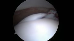
There are several modality options that may be recommended when diagnosing OCD (X-rays, CT scan, MRI, arthroscopy) (Figure 4). Commonly, diagnosis of OCD can be seen as a defect (flattening) present in the humeral head on X-rays of the shoulder. Despite apparent lameness in only one limb, radiographs of both shoulders are recommended because this condition may be present on both sides. Often, older dogs that have chronically untreated OCD present with large calcified fragments “joint mice” and osteoarthritis.
Seek veterinary advice if your young large breed dog is persistently lame in a forelimb, especially after exercise. Dogs with recurrent lameness unresponsive to medical treatment may require surgical intervention. Surgical treatment involves removal of the cartilage flap from the joint and scraping the edges of the bony defect to ensure removal of all affected cartilage. This may be accomplished through an open surgical or approach or arthroscopically. Arthroscopy is a minimally invasive procedure that uses very small portals through which a camera and specialized instruments can be passed to best accomplishes this task.
Your pet’s activity should be limited as directed by your primary care veterinarian in order to allow the incision(s) to heal. Gradually return your pet to full activity. Potential complications related to surgery include infection and postoperative seroma (fluid accumulation within the incision site) formation. Progressive osteoarthritis can occur with this condition, but is uncommonly associated with symptoms.


