A gallbladder mucocele is the distention of the gallbladder by an inappropriate accumulation of mucus.
Decreased bile flow, decreased gallbladder motility, and altered absorption of water from the gallbladder lumen are predisposing factors to biliary sludge. Biliary sludge may be a precipitating factor for the development of canine biliary mucoceles. However, it is more likely to be a small part of a complex disease process involving inflammation of the gallbladder wall and changes to the lining of the gallbladder changing the consistency of its secretions.
Hypersecretion of mucus leads to an accumulation of thick gelatinous bile within the gall bladder. Increased viscosity over a period of weeks or months leads to thick gelatinous material eventually occupying the entire lumen of the gallbladder and in some cases also being present in the ducts. The inciting cause of mucus hypersecretion is likely multifactorial and may be linked to certain diseases, such as:
- Cushing’s disease (hyperadrenocorticism)
- hypothyroidism
- inflammatory bowel disease
Certain genetic predispositions may play a role as Shetland sheepdogs were recently shown to be predisposed to gallbladder disease.
Clinical signs associated with gallbladder mucocele are often nonspecific and vague and, in some cases, a mucocele is discovered incidentally. Your pet may show signs including:
- decreased appetite
- anorexia
- lethargy
- vomiting
- diarrhea
- a yellowish tinge to the skin or gums
- abdominal pain or splinting
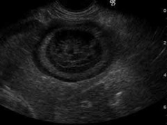
Diagnosis of the gallbladder mucoceles relies on physical examination by your primary care veterinarian and blood work combined with imaging modalities, like abdominal ultrasound. Abdominal ultrasound is incredibly useful early in the disease process and should be considered in any animal with clinical signs related to gastrointestinal upset (Figure 1). Up to half of pets with gallbladder mucoceles have a gallbladder rupture at the time of diagnosis and this number can be greatly reduced with early diagnostic intervention.
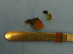
The presence and progression of gelatinous material in the gallbladder may lead to a life threatening obstruction of the bile duct or the inflammatory changes to the wall of the gallbladder can lead to rupture and spillage of bile contents into the abdomen (Figure 2).
A proactive course is recommended in most pets with gallbladder mucoceles. Pets with an incidental mucocele or “premucocele” on ultrasound should be considered for removal of the gallbladder, known as cholecystectomy. A current trend is to delay removal of the gallbladder of these patients until they have failed medical management, become systemically ill, or the gallbladder has ruptured. This “wait and see” philosophy may raise the likelihood of a life-threatening gallbladder rupture. Many surgeons are now recommending that gallbladder removal be performed in patients with gallbladder mucoceles at initial presentation or if found as an incidental finding on abdominal ultrasound. Routine open or laparoscopic cholecystectomy in the clinically unaffected gallbladder mucocele patient has been found to have an excellent outcome and rapid return to normal function in cases reported.
As with any surgical procedure, there are risks associated with general anesthesia. Preoperative blood work and imaging, and correction of fluid and electrolyte imbalances will help your ACVS board-certified veterinary surgeon minimize the anesthetic risk. Some patients may require intensive care support before and after surgery. Biliary surgery has inherent risks, including bleeding, and leakage of bile into the abdominal cavity, which can cause peritonitis. Abdominal drains may be needed to help manage peritonitis.
Your pet should be kept quiet for two weeks after gallbladder surgery, avoiding running, jumping, playing, stairs, or off-leash activity. The abdominal incision should be monitored for appropriate healing. Use of an Elizabethan Collar is often recommended to prevent licking or self-trauma. Medications for pain relief and to manage concurrent liver disease or infection are often sent home after surgery.
Dogs with gallbladder mucoceles that undergo cholecystectomy and survive the immediate perioperative period have an excellent long term prognosis. Overall mortality rates are reported to be between 20–39% for this disease, however, early surgical intervention may significantly reduce mortality rates. The resected gallbladder and a small piece of liver may be submitted for biopsy and bacterial culture by your veterinary surgeon. The results of these tests help the doctors caring for your pet to treat concurrent liver disease and infection.
Premier Sponsors


