An ectopic ureter is an abnormality of the ureter (the tubular tissue that connects the kidney to the urinary bladder) where the ureter does not enter into the urinary bladder in the correct anatomic position. This abnormality is something that cats and dogs are born with and can affect one or both of the ureters. The ectopic ureter may tunnel within the bladder tissue before opening in an abnormal location (intramural) or may implant into an abnormal area without tunneling (extramural). Additional congenital abnormalities of the kidneys and urinary tract can also be observed.
Symptoms of an ectopic ureter can occur as a young puppy or kitten or sometimes as a young adult. Female dogs are known to be 20X more likely to be diagnosed with ectopic ureters with certain breeds being more commonly affected including Golden and Labrador retrievers, Skye Terriers, etc. Symptoms can include:
- Continuous or intermittent urinary incontinence (leakage) or urinary accidents
- Difficulty potty training
- Urine leakage when laying down and/or sleeping
- Dribbling urine
- Recurrent UTIs
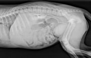
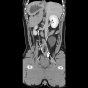
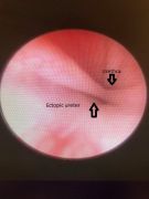
Your veterinarian may suspect ectopic ureters based on age, breed, and history. A full work up is recommended to rule out other causes of symptoms. This will likely include bloodwork to include complete blood count, chemistry panel, urine analysis and culture. Survey radiographs (x-rays) can be used to screen for stones or other abdominal abnormalities. An abdominal ultrasound can also screen for stones or bladder issues. In some cases, ectopic ureters can be seen on ultrasound. Advanced imaging is often required for a definitive diagnosis of ectopic ureters. This involves using intravenous dye with radiographs (Figure 1) or a computed tomography (CT) scan (Figure 2) to highlight the urinary tract and trace where the ureters are implanting. An endoscopic exam via vaginourethrograpy with cystoscopy is where a camera is introduced into the vagina, urethra and urinary bladder to evaluate the distal ureteral openings. Figure 3b Certain types of ectopic ureters may also be treated at the same time with this technique.
- Bloodwork: complete blood count, chemistry panel, urine analysis and culture
- Radiographs (x-rays) with contrast (special dye) given IV to help highlight the kidney and urinary tract. (Figure 1)
- Ultrasound to assess for anatomic abnormalities and to evaluate for any abnormal urine flow
- Computed tomography (CT) with contrast to evaluate the urinary tract. (Figure 2)
- Vaginourethrograpy with cystoscopy: a camera introduced into the vagina, urethra and urinary bladder to evaluate the distal ureteral openings. (Figure 3b)
Aftercare of these patients is dependent on the approach for treatment. If an open approach to the abdomen is performed then 2 weeks of activity restriction with incisional care is advised. If a minimally invasive approach using cystoscopy is performed then patients may return to normal activity much faster. Follow up serial urinalysis and cultures are advised
The main post-operative risks include continued incontinence, leakage of urine into the abdomen, stricture of the surgical site, and urinary tract infection. In some cases of continual incontinence further improvement may be achieved with incorporation of medications to help with urethral sphincter tone and/or the placement of an adjustable artificial urethral sphincter. Male patients with urinary incontinence secondary to ectopic ureters can have a higher success rates (70-80%) than females following surgical intervention. Unfortunately, even with surgical intervention and medical management 25-70% of female patients may still have persistent urinary incontinence.
Premier Sponsors


