A dermoid sinus, also known as a pilonidal sinus, is a tubular skin defect caused by incomplete separation of the skin and the nervous system during embryonic development. The sinus can be found at any point along the back or neck, but most commonly affects the neck or upper spine. The depth of the defect varies, the tube:
- may extend into the tissue just beneath the skin,
- may extend deeper and connect to the membrane covering the spinal cord (dura mater) or
- it may be a blind ended sac beneath the skin
Dermoid sinus is most common in Rhodesian ridgebacks, in which it is congenital and heritable. Therefore, affected pets should not be bred. The condition has also been reported in other breeds.
A dermoid sinus can be recognized at a young age as an opening on the midline of the back with protruding hair, often in a swirl. A tube or cord may be felt beneath the opening. Some dermoid sinuses may not be associated with any clinical signs or may be associated with mild discharge that can be controlled with gentle cleansing. However, sinuses that become plugged with keratin debris may become infected and an abscess may form. Sinuses that connect to the lining of the spinal cord can be associated with neurologic abnormalities.
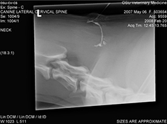
Figure 1
Your primary care veterinarian may probe the sinus by placing a catheter into the opening. A contrast fistulogram (Figure 1) can be performed by injecting contrast material into the sinus and then taking a radiograph. Because the depth of the sinus is often filled with hair or keratin debris, probing and contrast fistulography may underestimate the depth of the sinus. In this case or when connection to the lining of the spinal cord is suspected, advanced imaging may be recommended, such as:
- myelography (an x-ray test with contrast injected around the spinal cord)
- magnetic resonance imaging (MRI) or
- computed tomography (CT)
The prognosis for dogs with dermoid sinus is very good if no neurological signs are present. Failure to remove the sinus entirely results in redevelopment of the sinus which may require a second surgery to search for remnants of the tube that were left behind.
If surgical management involves the vertebral column at the cervical region, the most severe potential complication can be death due to sudden respiratory arrest with injury to the spinal cord during surgery. Other possible complications include failure of the repair with migration or breakage of the implants, inadequate reduction or mal-alignment of the spine. Implants can be positioned incorrectly causing chronic pain or impingement of the spinal cord, and requiring removal. Improper positioning can be a problem due to the small amount of bone available to engage the pins or screws, and a very small target area in small dogs to avoid the spinal canal.
Premier Sponsors


