Canine elbow dysplasia (ED) is a condition involving multiple developmental abnormalities of the elbow joint. The elbow joint is a
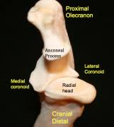
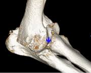
complex joint made up of 3 bones (radius, ulna, and humerus) (figure 1). If the 3 bones do not fit together perfectly due to growth abnormalities, abnormal weight distribution on areas of the joint occur causing pain, lameness, and the development of arthritis. Elbow dysplasia is a disease that encompasses several conditions grouped into medial compartment disease (fragmented coronoid process (FCP), osteochondrosis (OCD), joint incongruity, and cartilage anomaly) and ununited anconeal process (UAP). The cause of ED in dogs remains unclear. There are a number of theories as to the exact cause of the disease that include genetics, defects in cartilage growth, trauma, diet, and so on. It is most commonly suspected this is a multifactorial disease in which causes the growth disturbances, (Figure 1).
Elbow dysplasia is an inherited condition that can occur in most dog breeds but is most commonly seen in large to giant breed dogs. It has been noted to affect both elbows in up to 80% of patients. Bernese Mountain Dogs, German Shepherds, and Golden retrievers among others are predisposed to UAP while Labrador retrievers, German Shepherds, and Golden retrievers have an increased predilection among other breeds for developing medial compartment disease.
Unfortunately, once the elbow joint has been damaged through either cartilage loss, medial compartment disease or an ununited anconeal process, inflammation and further cartilage damage occurs. Ultimately this causes progressive arthritis of the elbow joint leading to pain and loss of function.
Dogs affected by elbow dysplasia often show signs from an early age, typically from 5 months on, but some may first be diagnosed after 4–6 years. Affected dogs develop a front limb lameness that typically worsens over a period of weeks to months. Lameness is usually worse after exercise and typically never completely resolves with rest. Often both fore legs are affected, which can make detection of lameness difficult, as the gait is not asymmetric. When both elbows are involved the dog usually becomes unwilling to exercise for long periods or may even refuse to complete a walk.
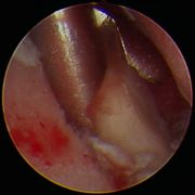
Diagnosis of elbow dysplasia is usually performed with a combination of clinical examination and x-rays. Usually the dog has pain on fully bending or extending the elbow and often your veterinarian will want to watch your dog walk or trot to detect any lameness. X-rays will typically shows signs of arthritis but may also show the presence of small bone fragments in the joint or an ununited anconeal process (Figures 2 and 3). Your veterinarian may also choose to refer you to a specialist veterinary surgeon for more advanced diagnostic procedures to be performed. This may include CT scans, MRI scans, or arthroscopy (Figures 4 and 5).
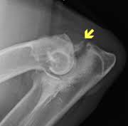
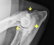
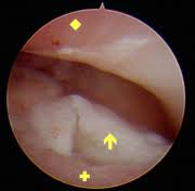
Figure 4: Arthroscopic image of a fracture coronoid fragment (arrow) with severe cartilage damage to the humerus (diamond) and ulna (cross).
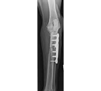
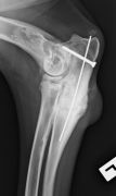
Treatment depends on the severity of the disease in the elbow. In most cases surgery is recommended, but your veterinarian may recommend medical management if the problem is very mild or so severe that the joint may not likely benefit from routine surgery. Treatment will depend on the primary cause of the elbow dysplasia. Often surgery is best performed arthroscopically, but conventional open surgery can also be done. Depending on the individual dog’s elbow problem surgery may involve:
- Removal of any coronoid fragments and removal of loose cartilage (Figure 6) (FCP).
- Surgical alteration of the elbow joint to shift weight away from damaged areas (Figure 7)
- Reattachment or removal of an united anconeal process (UAP) of the medial joint compartment (Figure 8)
- Correction of joint step/incongruity; this is usually done by cutting the ulna to re-establish elbow congruence.
- Joint replacement if the elbow is severely diseased
Surgery aftercare will depend on the type of surgery performed, and your veterinarian will advise you of exactly what is required. In general your dog will need to be quiet and confined for a period of time, usually from 2–6 weeks or more.
The outcome will vary between dogs, but in general the more mild the disease and the earlier it is treated, the better the long-term outcome. Most dogs will benefit from surgical treatment even if disease is more advanced, but unfortunately once arthritis is established it will slowly progress regardless of any treatment. On average, with treatment 85% of cases will show some degree of improvement in lameness and comfort despite progression of arthritis on x-rays. The aim of treatment is to slow the progression of arthritis and prolong the patients’ use of the elbow. Unfortunately elbow dysplasia cannot be cured but it can be well managed and our patients can have a good long term prognosis and outcome with a combination of surgical and medical management.
Premier Sponsors


