Anal sacs are paired small pouches located on either side of the anal opening in dogs, cats, ferrets and other animals. The lining of the sacs produces a smelly brown liquid that is usually eliminated in small quantities during defecation. The purpose of the anal sac is unknown but it is thought that they function in communication about the animal or its territory.
Anal sacs can become inflamed, infected, or impacted (blocked). With these conditions it is common for your pet to scoot their bottom on the ground and lick or chew at the area.
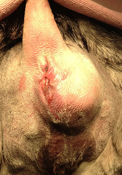
Tumors of the anal sacs (apocrine gland adenocarcinoma is the most common tumor type) are uncommon but can be a serious condition because they tend to invade surrounding tissues and metastasize (spread to distant tissues) even when the primary tumor is very small. The tumors are almost always only on one side. In approximately 25% of cases, the tumor can cause hypercalcemia (elevated blood calcium), which can cause kidney failure.
Anal sac tumors occur in male and female dogs with roughly an equal incidence. They occur in any breed but are more common in Spaniel breeds. Ten years is the average age of affected dogs. Anal sac tumors are very rare in cats.
The signs of anal sac tumors can be variable:
- an external swelling in the perianal region (Figure 1)
- a mass may be felt during a routine rectal examination
- constipation
- pain or straining to defecate
- blood in the stool
- excessive thirst and urination may occur if kidney failure is present from elevated calcium levels
- without enlarged lymph nodes (in the abdomen), even a large tumor often produces limited symptoms associated with defecation
Animal owners may observe symptoms of kidney failure from elevated calcium (referred to as hypercalcemia of malignancy).
These symptoms generally include:
- increased thirst
- increase in urination
- vomiting
- loss of appetite
- lethargy (weakness/tiredness)
In general, the following tests are recommended to diagnose the tumor, provide a clear clinical picture of overall health, and evaluate for metastasis:
- Needle Biopsy: A small needle is inserted into the tumor to obtain a few cells that can differentiate cancer from infection or inflammation.
- Blood tests: Assess overall health. Evaluating for hypercalcemia and kidney failure
- Chest x-rays: Evaluate for metastatic nodules and other heart and lung problems
- Abdominal ultrasound: Examination to evaluate for enlarged lymph nodes or tumor spread into other organs such as liver, kidneys, etc. These enlarged lymph nodes are often what produce symptoms associated with defecation.
Consultation with your primary care veterinarian may result in a referral to an ACVS board-certified surgeon to fully explore your options.
- Surgery is the mainstay of treatment. It is the only proven method to influence survival of dogs with these tumors. The tumor is removed through an incision near the anal opening directly over the tumor. Wide and aggressive removal is not possible due to the nearby important structures (rectum and anus). With large tumors, additional tissue attached to the tumor may need to be removed. This may result in some of the complications discussed below.
- If there are enlarged lymph nodes in the abdomen, they are removed through an abdominal surgical approach on the underside of the dog. These nodes are enlarged in about 50% of cases. This can be done at the time of the primary tumor removal, shortly thereafter, or later if these nodes enlarge. This procedure is done to alleviate constipation and difficulty defecating.
- If kidney failure or hypercalcemia is present, therapy with intravenous fluids and medications may be needed prior to surgery to make your dog a more suitable candidate for anesthesia. In some cases, kidney failure can be permanent.
- After surgery, chemotherapy, and/or radiation treatment may improve the life expectancy of your pet.
Most animals are discharged 1-2 days after surgery. There is usually a follow-up appointment to see how your dog is doing and to remove skin sutures or staples (if present). Pain can be well-controlled with oral medications.
Restrictions following surgery usually are:
- use of a restrictive collar for 10-14 days after surgery to prevent the natural tendency of dogs to lick and chew at a wound. This can cause breakdown of the wound and infection.
- stool softening medications may be needed until swelling resolves
- limited and restricted activity is indicated for about 2 weeks to allow recovery and incision healing
Postoperative complications can include:
- incision infection
- wound breakdown (dehiscence)
- fecal incontinence can occur in up to 33% of dogs especially with removal of larger masses. This is usually temporary but owners need to be aware of this problem. If the tumor is only on one side, the incontinence is typically partial in that the dog has difficulty controlling bowel movements but not continuous dropping of stool
- continued kidney problems
The prognosis with apocrine gland adenocarcinoma depends on type of treatment, size of mass, presence of hypercalcemia, and presence of lymph node involvement. Surgical removal of these nodes can produce long-term relief of constipation. Some animals have had multiple surgeries to remove recurrent lymph nodes to alleviate obstructions successfully. Your veterinarian will likely recommend follow-up with a veterinary oncologist after your pet heals from surgery to discuss other treatments for long-term success for dogs with this disease.
It is important that your veterinarian examine the anal sacs as part of your dog’s routine examination. Early detection can greatly improve survival.
Premier Sponsors


