
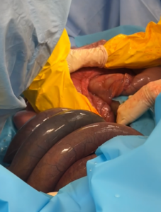
With colic, several conditions can damage the intestines and require surgery. The small intestine, which connects the stomach to the cecum, is a narrow but lengthy part of the digestive tract. In horses, it is typically 50–70 feet long, making it more likely to become twisted or trapped.
Some conditions can cut off the blood supply to a portion of the small intestine. When this happens, the affected tissue can die and must be surgically removed. Other problems, such as adhesions (bands of tissue that cause parts of the intestine or other organs to stick together) or masses, may also require removal of the affected section.
Removing a portion of the intestine is called a resection. Reconnecting the remaining ends of the intestine is called an anastomosis.
A horse with a problem that requires small intestinal resection will usually have severe colic signs, but once the tissue dies, they can appear comfortable. Different ages and breeds of horses show pain in different ways, and some horses will have milder signs if they have a high pain tolerance.
Typical signs associated with colic are often observed such as:
- Poor or no appetite
- Occasional or constant signs of colic (kicking, pawing, rolling, flank-watching)
- Decreased or no manure production
As the intestine becomes more compromised, these signs may indicate a more severe problem and occur within minutes to hours:
- Purple/dark gums
- Lethargy/listlessness
- Trembling
- Bloated appearance (abdominal distention)
- Shock-cold limbs, nose and extremities along with gum color changes
If a portion of the intestine dies within the abdomen, and it is left untreated, the horse will usually die within a day.
A full colic workup is often performed, starting with:
- Perform a physical examination, including checking the horse’s heart rate, temperature, and respiratory rate; listening to the abdomen and chest; and examining the mucous membranes.
- Nasogastric intubation: A tube may be passed up to the horse’s nose to check for reflux, which can provide the horse with relief if there is pressure in the stomach or reflux (vomit). Frequently a horse with a small intestinal lesion requiring resection will have a large volume of reflux, but it can take time to build over the course of the colic episode.
- Rectal examination: Palpation of distended loops of small intestine is possible in the majority of cases needed for a small intestinal resection; not being able to feel distended loops does not necessarily mean that a small intestinal problem can be ruled out.
- Ultrasound examination: Distended loops of small intestine, without normal GI movement (amotile), can be seen ultrasonographically and often indicate a problem requiring surgery.
- Blood work: Dehydration is common; an elevated lactate can indicate that the small intestine is not getting enough blood flow. Electrolyte imbalances are also common due to the small intestine not functioning properly.
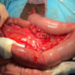
Small intestinal resection is performed during abdominal surgery. The piece of intestine that needs to be removed separated from the other intestines and organs to avoid spillage into the abdomen and to maintain sterility. The blood vessels going to that part are tied off, and the segment is cut away from the neighboring intestinal segments. The healthy intestine pieces are then sutured together and the intestine is checked to make sure that food can pass through the sutured part without leaking.
Aftercare can vary widely depending on how much intestine is removed and how bad the damage is before it is removed. In most cases, the following treatments are used:
- IV fluids
- Pain medication
- Anti-inflammatory medication
- Slow refeeding or a low-residue(senior feed) diet
- Stall rest-some clinicians may recommend short periods of handwalking depending on the case
More severe cases may require:
- Motility drugs
- Anti-endotoxin medication such as polymyxin B
- Ice boots
- Abdominal lavage with a drain after surgery
Each medical team will decide what is best for your horse at the time.
After hospitalization for a few to several days, most horses will be discharged to continue recovery at home. It is important to closely follow your surgeon’s recommendations regarding exercise and feeding. Some horses will be at risk of adhesions after surgery which can cause repeat colic. In most cases, the horse will need a period of strict stall rest followed by small pen or pasture turnout with no forced exercise for 3 months. Some horses may also need bandaging to support their incision as they heal, but it is important to follow your provider’s recommendations for you and your horse.
Premier Sponsors