Horses have a very well developed “fight or flight” mechanism and they often react first and think later. If they are caught or cast against something such as a fence or barn wall, their first instinct is to flee; often without regard to whatever body part happens to be caught at the time. Therefore, horses have a well-deserved reputation as being accident-prone.
Depending upon the wound location and the amount of time since it first occurred, you might see some of the following signs:
- Lameness or unwillingness to bear weight on the affected leg and/or unwillingness to come in from the pasture or move from where the horse is located.
- An area of dried blood or an accumulation of flies to a small wound
- Flap of skin hanging off
- Profuse bleeding
- Swelling or puffiness of the surrounding area
- Heat associated with the swelling
Primary reasons to seek your primary care veterinarian’s assistance:
- Excessive bleeding
- Significant contamination with dirt/debris
- Lameness
- Full-thickness wounds (the skin has been cut) or wounds involving deeper structures such as bone, muscle, joints, ligaments, tendons, tendon sheaths, vessels, and nerves
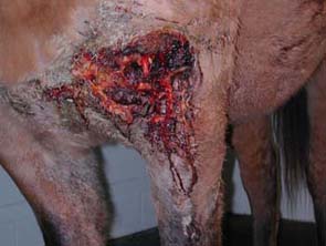
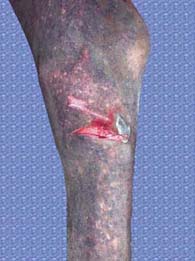
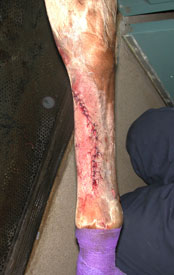
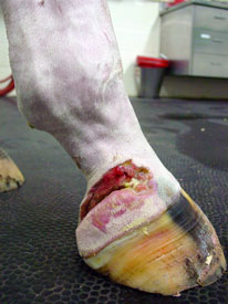
Call your veterinarian to evaluate any wound that your horse may have. Often times the smallest puncture wound may cause a more serious life-threatening injury when compared to a very large wound. A very large wound on the upper body that is not close to joints and is not very deep may have a much better prognosis than a small puncture wound that may look less severe on the lower limb into a tendon sheath or joint (Figures 1 and 2).
Your veterinarian may perform the following procedures to evaluate your horse:
- Physical examination
- Thorough examination of the wound itself
- Wound exploration to determine the depth and width of its borders, removal of any foreign bodies such as wood, wire, pieces of bone or hair, and to determine if any vessels, nerves, tendons, ligaments, muscle, bone, or joints are involved
- Thorough examination may require local anesthesia and sedation. In some cases, horses may require general anesthesia to fully assess the severity or complexity of the wound
- Radiographs of the area with or without a contrast agent
- Ultrasound examination to evaluate the soft tissues, nearby joints, and locate potential foreign bodies
- A sample of joint or tendon sheath synovial fluid, if involvement is suspected
- Involvement of a joint or synovial sheath significantly complicates the wound healing process and can lead to an infected joint. Repeated lavage or flushing of the synovial structure is indicated and may or may not be curative.
Immediate treatment you can provide while waiting for your veterinarian to arrive:
- Application of direct pressure to any area that is actively bleeding with an absorbent pad (ex: sheet cotton, shipping bandage quilt, folded bath towel, or several layers of roll cotton) covered with an elastic bandage material (Elastikon, VetWrap etc.)
- Move your horse into a clean, quiet environment while waiting for your veterinarian to arrive, if possible
Your veterinarian may provide the following treatments:
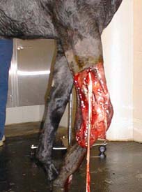
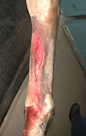
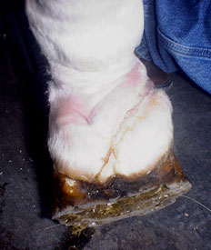
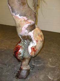
- Thorough clipping, cleaning, and removal of any visible foreign matter (Figure 4a, 5a)
- Removal (debridement) of damaged or contaminated tissue of the wound
- Suture closure, if appropriate & possible (Figure 4b)
- Bandaging or casting, depending on location (Figure 5b)
- Administration of broad spectrum systemic antibiotics and/or anti-inflammatory medications
Following initial stabilization and care of the wound, your veterinarian may recommend that your horse be restricted to stall rest +/- hand-walking, but the extent and location of the wound will dictate the level of exercise.
Several factors can affect outcome:
- Location:
- Head – Involvement of the sinuses, nasal passageway, eye etc. Wounds to this area heal relatively well as long as critical structures are not involved
- Upper limbs – Have skin/muscle covering, but motion in these areas can negatively affect healing
- Head and upper body wounds have a relatively good blood supply compared to lower limb wounds
- Abdomen – If the wound is deep enough, it could penetrate the abdomen and cause peritonitis (infection of the abdominal cavity)
- Upper body and abdominal wounds have sufficient skin and underlying muscle to allow significant wound contracture to help close even very large defects compared to wounds on the lower limbs
- Lower limbs – have only skin covering very important structures such as bone, tendon, ligament, joints, tendon sheaths, vessels, and nerves
- Over a joint – Movement of the skin/wound over this area can negatively affect healing
- Wounds below the carpus (knee), hock or over a joint are complicated by minimal excess skin that is fairly inelastic, lacks underlying muscle tissue, and is subject to movement during limb flexion. These wounds are also prone to exuberant granulation tissue (“proud flesh”), which will dramatically slow healing and may require surgical intervention
- Contamination:
- Dirt, manure, hair, or other foreign material: Can infect the site or underlying structures & may be life threatening if infection is not resolved
Complications of the wound healing process include:
- Cellulitis
- Subcutaneous emphysema (air under the skin)
- Hematomas/Seromas (accumulation of blood/serum)
- Sarcoids or squamous cell carcinoma tumors
- Exuberant granulation tissue (proud flesh) (Figure 6)
- Prolonged healing
- Excessive scarring
- Laminitis
- Colic
If joints or tendons are involved, additional complications include:
- Persistent infection that is resistant to treatment and can be life threatening
- Development of arthritis
- Adhesions of tendons
- Persistent lameness
- Restriction of motion due to excessive scar tissue
After evaluation and continued care by your veterinarian of your horses wound, they will be able to discuss with you the specifics related to overall prognosis.
Premier Sponsors