
Subchondral cystic lesions (SCLs), also known as subchondral lucencies or subchondral bone cysts, are fluid-filled lesions within the area underneath the joint cartilage surface, which called subchondral (or under the cartilage) bone. SCLs can be developmental lesions and are identified in young (1-3 years of age) horses, or can result secondary to joint injury or osteoarthritis, which are typically diagnosed in adult horses (over 5 years of age). While they most commonly occur under the joint surface, they can also occur next to growth plates. SCLs most commonly occur in the medial femoral condyle within the stifle joint, particularly as a developmental lesion in young horses. Other sites include bones of the pastern joint, the navicular bone, and the radius, among other sites.
Symptoms of SCLs can vary widely depending on factors such as the size and orientation of the cyst, the age of the horse, and whether it is developmental or secondary to injury or osteoarthritis. Some cysts are identified on routine radiographs, often performed evaluating for developmental lesions or pre-sales. Often horses show mild to moderate lameness which may wax and wane. Typically, they are accompanied by joint effusion, or extra fluid within the joint. Particularly in young horses’ cysts can occur in multiple joints, even if clinical signs are primarily present in one joint, so a thorough examination +/- imaging may be warranted to evaluate for multifocal disease.
Lameness evaluation, joint palpation, and local anesthesia through nerve and joint blocks can often localize the site where lameness is originating. Most SCLs can be identified through radiographs; however, some require computed tomography (CT scan) to fully define the extents of a cyst and any additional lesions within a joint. Additionally, nuclear scintigraphy (bone scan) may be useful for cysts at less common locations. Ultrasound may also be a useful imaging modality to determine whether the cartilage overlying the cyst appears intact or abnormal.
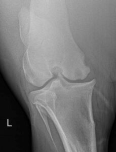
A variety of non-surgical and surgical management options are available and will depend on the size of the cyst, communication with the joint, age of the horse, the presence of concurrent joint disease, long-term goals for the patient, and several other factors. Only a conversation between you and your veterinarian can determine the ideal course of treatment. However, some of the options are listed below:
- Wait and watch: There is evidence that small lesions in young horses may spontaneously resolve, and some lesions may warrant repeat imaging at a specified time following diagnosis to determine whether additional treatment is warranted.
- Rest +/- Nonsteroidal anti-inflammatory medications: Rest and non-steroidal anti-inflammatory medications as needed may allow improvement of small lesions in young horses. Success rates vary widely in the literature (45-65%). However, there are reports of cyst recurrence following conservative management as well, and this should be kept in mind when considering treatment options.
- Corticosteroid injection: The goal of corticosteroid injection is to reduce inflammation associated with the cyst. While the joint itself can be injected with corticosteroids, improved success occurs with intralesional injection, which typically needs to be performed under anesthesia either using arthroscopic or ultrasound guidance to direct the needle directly into the cyst.
- Cyst debridement: Debridement involves evaluating the joint arthroscopically, using small incisions to pass a camera and instruments into the affected joint, then removing the abnormal cystic tissue. This will create a defect, which can be packed with a variety of materials to stimulate healing.
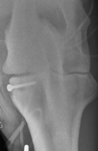
- Translesional or transcondylar cortical screw: This technique has been described across several anatomical locations. Application of a screw across the lesion or portion of bone affected compresses the cyst area, inducing bone formation within the cyst lesion. (Figure 2)
Aftercare will depend upon the decided course of treatment, but for most a period of rest will be warranted. Surgical procedures will have incisions that require monitoring and likely suture removal following healing. Any procedures involving entering the joint (injection or surgery) carry a risk of infection, and any concerns should be brought to the attention of the referring veterinarian or surgeon.
Outcome depends on the size of the cyst, the size of cartilage lesion, the age of the horse, cyst location, and whether osteoarthritis is present in the joint. Reported success ranges from 50-75% for athletic use and soundness. For cysts large enough to warrant intervention (through injection or surgery) intervening in younger horses (less than 3 years old) carries a better prognosis than waiting until the horse is older, when secondary osteoarthritis is more likely to develop.
Take-home: Subchondral cystic lesions are a moderately common developmental orthopedic disease in young horses, but with appropriate care many of these horses can have long productive athletic careers. There are several treatment options, and picking the best course depends on several patient, owner, and lesion specific factors. Ultimately working with an ACVS board certified surgeon along with your referring veterinarian will bring the best team to the table to get your horse happy, healthy, and sound again.
Premier Sponsors