
The small metacarpal and metatarsal bones in the horse are vestigial structures that are located on the inside and outside of each cannon bone and are commonly referred to as “splint bones”. Because of their location these bones are prone to fracture from external trauma such as a kick from another horse. However, splint bone fractures can also occur during exercise and may occur secondary to injury of a soft tissue structure such as the suspensory ligament.
Splint bone fractures are classified by the location of the fracture within the bone (upper (proximal) third, middle third, and lower (distal) third). In addition, these fractures can be simple or comminuted (multiple fragments) and when trauma is the cause they often present with an associated wound. Treatment recommendations are based on classification of the fracture.
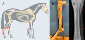
Horses present with varying degrees of lameness depending on the length of time between occurrence of the injury and the location of the fracture. Lameness is typically more severe at the onset of injury but can be more subtle for chronic injuries. Acute fractures of the upper third of the splint bone often show lameness at the walk whereas fractures lower in the bone may only show lameness at the jog or trot.
Swelling, heat, and pain on palpation at the site of the fracture and the surrounding soft tissues is common, and a wound may be present. Early in the injury the swelling is soft to firm, but over time the swelling may become hard due to callus formation at the fracture site. Fractures can occur at the medial (inside) or lateral (outside) splint bone although fractures from trauma more commonly occur at the lateral splint bone due to its location on the outside of the limb.
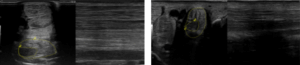
Radiographs (x-rays) confirm the diagnosis of a splint bone fracture, but ultrasound is often recommended to evaluate the surrounding soft tissue structures especially the suspensory ligament. Computed tomography (CT) can be useful in chronic fractures with signs of infection or sequestrum formation and in cases that may involve the nearby cannon bone.
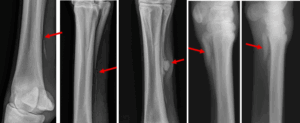
Treatment of splint bone fractures depends on the location of the fracture and complicating factors such as a wound. Depending on the type of injury, some splint bone fractures can be managed conservatively while others require surgical treatment. Treatment decisions should be made by the primary care veterinarian and/or an ACVS board-certified veterinary surgeon in conjunction with the owner.
- Conservative therapy: non-surgical management typically includes stall rest for four to six weeks. If a wound is present, wound care and treatment with antibiotics may be indicated. Fractures of the upper portion of the splint bone with a corresponding wound are good candidates for conservative therapy since placement of surgical implants in the face of an open wound are at high risk of becoming infected. Other types of fractures that are good candidates for non-surgical management include fractures that are minimally displaced (bone fragments maintain good alignment) and acute fractures.
- Surgical management: the most common surgical treatment options for splint bone fractures include removal of the fractured portion of the splint or internal fixation with a bone plate and screws.
Removal of the fragment is often performed under general anesthesia although some veterinary surgeons may prefer standing procedures. Up to two thirds of the splint bone can be removed safely without any detriment to soundness. Therefore, removal is typically the recommended surgical treatment for all fractures that involve the lower two thirds of the bone.
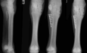
Internal fixation, or fracture repair, of proximal splint bone fractures is reserved for cases that are not amenable to conservative therapy and are located in the upper third of the splint bone. The fracture is repaired with a small plate and screws treatment of these types of fractures with screws alone has also been reported but has a high complication rate.
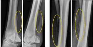
Fractures treated with conservative therapy typically require 8-12 weeks of controlled rest before allowing turnout or ridden exercise. It is important to monitor healing with radiographs because if the fracture is not healing as expected then surgical therapy may be indicated.
Splint bone fractures treated with removal have a shorter convalescence period as long as there is no corresponding suspensory ligament injury. After removal, horses are rested and maintained in a bandage for two weeks until suture removal and then kept in a standing wrap as needed for swelling. Horses are often back in work by four to six weeks after surgery.
Fractures treated with internal fixation are kept on a controlled rest and rehabilitation program, with return to work, barring complications in three to four months. Post surgery these horses are also kept in a bandage for two to four weeks and may be treated with post-operative antibiotics due to increased risk of infection due to the implants.
Prognosis for return to athletic function with conservative therapy is very good as long as the fracture shows adequate healing with little callus formation. Excessive callus formation can impinge on the suspensory ligament and cause long term soundness issues. Horses that treated with splint bone removal have an excellent prognosis for return to their previous level of exercise if there is no concurrent suspensory ligament desmitis. Prognosis for fixation with plates and screws is good with a recent report showing a 76% return to the horse’s previous level of work.
Premier Sponsors