Tibial growth plate fractures are injuries that occur in the growing bones of young dogs. The tibia, or shinbone, has areas near the ends called growth plates that allow the bone to lengthen as the puppy grows. These areas of soft developing bone close as they mature. However, they are softer and more vulnerable to injury than mature bone. Most tibial growth plate fractures are associated with minimal trauma, such as a fall, getting a leg caught, or simply rough-housing with another dog.
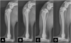
Common types of fractures near the growth plate of the tibia in young dogs include:
• Tibial tuberosity avulsion fractures, where a small piece of bone (the tibial tuberosity) gets pulled off (Figure 1B)
• Proximal tibial physeal fractures, which involve the main growth plate of the tibia (Figure 1C)
• Proximal tibial metaphyseal fractures, involving a bigger part of bone lower than the growth plate (Figure 1D)
These injuries are most common in puppies 4 to 8 months old, especially in medium to large breeds with high activity levels.
You may notice:
- Sudden lameness or limping of a back leg
- Swelling near the knee
- Pain or inability to extend the leg
To diagnose these fractures, your veterinarian will:
- Perform a physical exam
- Take X-rays to assess the location and severity of the fracture. Sometimes, X-rays of the opposite leg are also taken to compare the normal growth plate appearance
Initial Management
After the injury and before you get to a veterinary clinic, you should confine your dog to a very small space, ideally a box or kennel. Movement should be limited to only necessary, such as to go to the bathroom. Seek veterinary care as soon as possible.
Fracture repair
For treating tibial growth plate fractures, surgery is usually necessary, especially when the fracture segment is fully detached or with significant displacement. Repair of the fracture involves fracture reduction (placing the fractured segment back into place), and stabilization with implants.
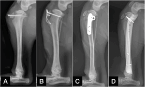
Commonly used implants in tibial growth plate fractures include orthopaedic pins (Figure 2A), wires (Figure 2B), and sometimes bone plate and screws (Figure 2C) or external fixator (Figure 2D).
In cases when the fracture displacement is small and in small-breed dogs, cast or splint may be considered.
Strict activity restriction is required during the healing period. Confinement often includes restricting your dog in a kennel or a small section of the house in the pen. Use rugs or yoga mats to cover slippery floors. Do not allow jumping on/off couch or bed. Also, do not allow running, jumping, or playing with other dogs. The only allowed activity should be doing out for bathroom breaks and always on a short leash. Most dogs start to use the injured leg 1-2 weeks after surgery. However, this does NOT mean the fracture is finished with healing. With the fracture stabilized with implants, your dog will want to use the leg, but can often overuse the leg during this healing period when not being restricted. The restriction should continue until bone healing is confirmed with X-rays. Too much activity too early can lead to implant breakage or migration, resulting in the need for second surgical repair.
Follow-up X-rays are usually done 4–6 weeks after surgery to confirm bone healing and implant positions. In some dogs, the implants may be removed once the bone has healed. After the follow-up exam, your veterinary surgeon can determine if your dog is ready to return to function.
The prognosis is generally good with surgical reduction and repair. With early treatment and proper healing, most puppies return to normal function and activity.
Premier Sponsors


