Mammary (or breast) tumors are common in female dogs, but rare in male dogs and cats. Surgical removal is recommended for most mammary tumors. Chemotherapy may be required following surgery in some cases. The prognosis is good following surgical resection for most mammary tumors in female dogs, but the prognosis is worse for certain types of tumors in dogs and all mammary tumors in cats.
Of dogs, poodles, dachshunds, and spaniels are most affected. In cats, Siamese or other Oriental breeds and domestic short hairs are most often affected. In dogs, obesity at a young age is a risk factor.
Mammary tumors are more common in female dogs that are either not spayed or were spayed after 2 years of age. The risk of a dog developing a mammary tumor is 0.5% if spayed before their first heat (approximately 6 months of age), 8% after their first heat, and 26% after their second heat. Cats spayed before 6 months of age have a 7-times reduced risk of developing mammary cancer and spaying at any age reduces the risk of mammary tumors by 40% to 60% in cats.
More than a quarter of unspayed female dogs will develop a mammary tumor during their lifetime. The risk is much lower for spayed female dogs, male dogs, and cats of either gender. In female dogs, 50% of mammary tumors are benign and 50% are malignant. However, few of the malignant mammary tumors are fatal. In contrast, over 85% of mammary tumors in cats are malignant and most of these have an aggressive biologic behavior (i.e., mammary tumors in cats tend to be locally invasive and spread elsewhere in the body).
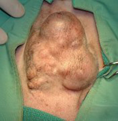
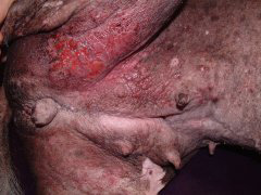
A palpable mass underneath the skin of the abdomen or near a nipple is the most common findings in dogs and cats with mammary tumors (Figure 1). However, other signs and symptoms include discharge from a mammary gland, ulceration of the skin over a gland, painful, swollen breasts (Figure 2), loss of appetite, weight loss, and generalized weakness.


A good general physical exam is needed to find the location, size, and character of all the mammary masses and assess local lymph node enlargement. Other procedures are performed to stage the cancer (determine what type it is and where it is located in the body):
- Bloodwork: blood count, chemistry, urinalysis, and clotting profile
- Abdominal ultrasound, chest x-rays and sometimes CT scan: used to check for cancer that has spread to other parts of the body
- Aspiration (aka Fine Needle Aspirate): a needle is used to sample the mammary mass to help distinguish it from other skin tumors. Lymph nodes may also be assessed to look for a spread of cancer cells. This is more reliable in dogs than cats to confirm a diagnosis.
- A biopsy may be indicated to rule out a certain kind of tumor called inflammatory mammary carcinoma, as surgery is generally not recommended for this tumor type

Further consultation with your primary care veterinarian may result in a referral to an ACVS board-certified veterinary surgeon to fully explore your options.
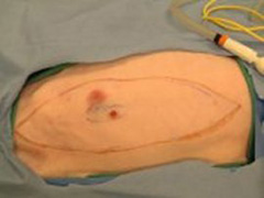
- The type of surgery depends on the size, location, and number of mammary tumors and species of your pet. In general, surgery is more conservative for dogs with mammary tumors and involves removal of either the mass alone or the affected mammary gland. However, in cats, more aggressive surgery is recommended with removal of one or preferably both sets of mammary glands (Figure 3). Surgery to remove both sets of mammary glands from a cat is often done in two surgical procedures 2–3 weeks apart.
- The local lymph node should also be removed in cats, if possible, to assess for evidence of spread of the cancer.
- The role of spaying female dogs with mammary tumors is controversial. Most studies have shown no beneficial effect of spaying at the time of tumor removal in preventing the development of new mammary tumors, or influencing the aggressiveness or metastatic potential of existing mammary tumors. However spaying at the time of mammary tumor resection may be recommended by your veterinary surgeon because recent studies have shown a beneficial effect in dogs with mammary tumors, and it can prevent unrelated diseases, such as pyometra (uterine infection).
- The role of chemotherapy in cats and dogs with malignant mammary tumors has not been clearly defined for all tumor types but oncologist consultation after surgery is often recommended. For most mammary tumors in cats and dogs, hormonal therapy, immunotherapy, and radiation therapy have either not been investigated or are not beneficial.
Surgery is not recommended for dogs with inflammatory mammary carcinoma because it does not improve survival rate. Unfortunately, an effective treatment has not been discovered. Radiation therapy in combination with a non-steroidal anti-inflammatory drug has been shown to provide the most effective pain relief in dogs, but the prognosis remains poor.
Most pets are discharged 1–5 days after surgery, depending on their extent of surgery and their comfort. They are usually returned for re-check and removal of skin sutures or staples (if present). Pain can be well-controlled with owner-administered medications. Pain patches may be adhered to the skin. Antibiotics may be sent home with some patients.
Restrictions following surgery usually are:
- Elizabethan collar for 10–14 days after surgery to prevent the natural tendency of pets to lick and chew at a wound.
- Limited and restricted activity is indicated for about 2 weeks to allow recovery and incision healing.
- Bandage care may also be required if one is applied.
- Kitty litter made from newspaper may be recommended rather than clay litter.
Postoperative complications can include:
- Incision infection
- Incision opening or breakdown (called dehiscence), which is more common in the mammary glands near the back legs or when larger areas of tissue have been removed
- Local recurrence of the tumor or spread of the cancer that was not detected at the time of surgery
If the mammary tumor is malignant, the surgical site and regional lymph nodes should be checked for local tumor recurrence and metastasis, respectively, every 3 months for the first 12 months after surgery and then every 6 months thereafter. Abdominal ultrasound and chest radiographs are also recommended every 3–6 months to assess for evidence of metastatic disease.
In dogs, there are a number of factors that influence the prognosis following surgery. These prognostic factors include tumor size, clinical stage (how far the cancer has spread in the body), tumor type and grade, and various other pathologic changes seen in the tumor tissue. Benign tumors are cured by surgery, although the development of new mammary tumors (both benign and malignant) is possible. There is a poorer prognosis with malignant mammary tumors and it also depends on what type of cancer. In dogs, the size of malignant mammary tumors is an important consideration when determining prognosis, both for local tumor recurrence and survival time. The smaller the mass is at the time of surgery (3–5cm or smaller) the less likely it will recur, or metastasize (spread) elsewhere. Dogs can live several years after complete removal of some malignant mammary tumors. So once a mass is found, having surgery to remove it earlier is better.
The prognosis for cats with mammary tumors is guarded as mammary tumors tend to be more aggressive and metastatic in cats. Many of the prognostic factors used in dogs also apply to cats, although the extent of surgery is also important in cats. When the tumor is smaller (less than 3cm) when it is removed, cats can live a couple of years; if it is larger than 3cm the prognosis for survival time after surgery is 4–12 months. Therefore, early surgical treatment is important. In cats, the amount of surrounding tissue removed with the mass can also affect how long they live after surgery. In some cases, survival time after surgery have been tripled in patients treated aggressively with surgery -those with large resections or tumor removal with wide margins lived 3 years and those treated with only tumor removal in the same tumor type lived 1 year.
Mammary tumors can be largely prevented by spaying before 6 months of age or before your pet’s first heat cycle. Other factors that may reduce the incidence of mammary tumors include feeding a well-balanced diet, avoiding obesity and avoiding the administration of hormones (particularly progesterone or mixed estrogen-progesterone drugs).


