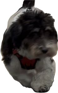
If your pet’s front leg looks “crooked,” “bowed,” or the paw points outward/inward, your pet may have Angular Limb Deformity (ALD)—most commonly affecting the radius and ulna (the two forearm bones) (see Figure 1).
ALD is a condition where a growing bone—most often the radius in the forelimb—develops an abnormal curve, angle, or twist. The good news: with careful planning and modern orthopedic techniques, your pet can usually be safely and accurately treated—and return to an active, comfortable life. This article focuses on front limbs; however, the principles of diagnosis and treatment often apply to both front and back limbs.
- ALD can occur when a single bone growth plate becomes diseased and results in a reduced growth potential. When only a single growth plate is affected, the surrounding bones continue to grow and the disparity of growth between associated bones can then result in slow deformity of a limb (curve, angle, or twist).
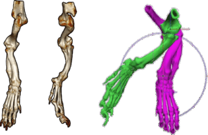
- Modern imaging (X‑rays and CT) and planning—including 3D‑printed models and patient‑specific cutting guides—allow your ACVS board-certified veterinary surgeon to straighten the limb with precision (Figures 3–4).
Treatment decisions are individualized and should be made by you and your ACVS board-certified surgeon together based on your pet’s needs and goals.
What is ALD and why does it happen?
In dogs, the radius and ulna grow alongside each other. If one growth plate is injured or closes too early, the other bone will continue to grow, but due to the halted growth of a single bone will then create forces that can bend, angle, or rotate the limb. Over time, this can strain nearby joints (elbow, wrist/carpus, shoulder) and contribute to discomfort or arthritis if not addressed.
- A visibly “crooked” or “C‑shaped” forelimb
- Paw or toes pointing outward or inward
- Limping after play, stiffness, or reluctance to jump/run
- Uneven nail wear, paw scuffing, or an abnormal gait
- In advanced cases, difficulty bearing weight or signs of persistent pain
These changes often appear in growing dogs (commonly 5–12 months), especially small and medium breeds, but any dog can be affected. Early evaluation helps simplify correction and improve outcomes.
Physical exam: Your ACVS board-certified veterinary surgeon will assesses limb alignment, joint motion, gait, and checks the elbow,
wrist (carpus), and shoulder for soreness.
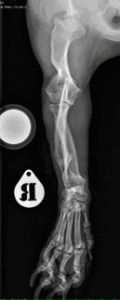
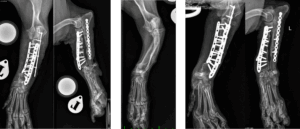
X‑rays (radiographs): Two standard views are typical, though more views may be taken per limb to measure where and how much the limb is bowed or angled (Figure 2).
CT scan: Provides 3D detail—especially helpful when the limb is both angled and twisted (torsed). CT improves the accuracy of surgical planning (Figure 3).
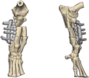
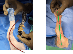
CT images can also be used to create 3D patient-specific bone models and cutting guides that can help your surgeon perform a precise correction and may shorten surgery time (Figure 4).
Treatment options pursued will depend upon your pet’s comfort and clinical severity. Pets with minimal deformity and are not affected by this anatomic change can be monitored, especially if your pet is still growing. Your veterinarian may recommend activity adjustments and weight management to reduce joint strain and help reduce discomfort associated with arthritis.
When the deformity causes pain, lameness, or significant misalignment, surgery—called a corrective osteotomy (one or more planned cuts) or a corrective ostectomy (removal of a small section of bone at a precise location)—may be recommended. The goal is to realign the bone into a straighter, more functional position and stabilize it, most often with a plate and screws sized for small animals (Figures 4–6).
- What can be corrected: side‑to‑side angulation (varus/valgus), forward/backward curve (procurvatum/recurvatum), and rotation (torsion).
- How the bone is stabilized: typically with a small‑animal plate and screws. In some cases, the ulna is also addressed to relieve tethering and reduce the chance of the deformity recurring.
- Why technology helps: modern locking plates and digital 3D planning with patient‑specific guides improve precision and efficiency.
- Important note: There are multiple acceptable techniques. The best option is individualized by your ACVS board-certified veterinary surgeon for your pet’s age, severity and type of deformity, comfort, and your goals.
- Pain control: Your pet will receive multimodal pain relief during and after surgery; you’ll go home with medications and clear instructions.
- Activity: Strict rest at first, followed by a gradual, supervised return to activity over about 8–12 weeks.
- Rechecks: Scheduled visits and X‑rays confirm bone healing and implant position.
- Rehabilitation: Gentle, guided exercises help restore strength, comfort, and a normal gait.
- Outlook: Most pets show improved comfort and limb use within weeks. Correcting alignment reduces stress on nearby joints and may lower long‑term arthritis risk.
- Risks: Infection, implant issues, stiffness, or under/over‑correction are uncommon but possible. Careful planning, precise technique, and following aftercare instructions help reduce these risks.
Premier Sponsors


