Young foals can commonly have crooked legs (either front, hind or both), otherwise known as Angular Limb Deviation or Deformity (ALD).
Causes:
- Perinatal factors: premature birth, twin pregnancy, placentitis, perinatal soft tissue trauma and flaccidity or laxity of the soft tissue structures surrounding the joints
- Developmental factors: unbalanced nutrition, excessive exercise and/or trauma
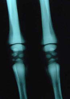
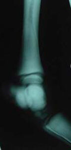
Most foals will be born with some degree of limb deviation, mostly due to ligament laxity and muscle weakness, which will usually correct itself as the foal exercises. If the cuboidal bones are not fully formed (Figures 1a and 1b), there is risk of the incompletely formed bones being crushed from exercise and uneven load that is placed on the joint due to the laxity. In these cases, once the bones have fully formed/calcified, an ALD will result due to the abnormally shaped cuboidal bones.
There are two general terms used to describe these deviations:
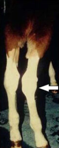
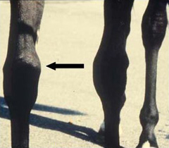
- Valgus refers to an outward deviation of the limb (Figure 2).
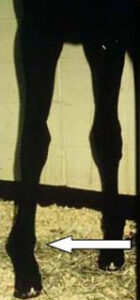
- Varus refers to inward deviation of the limb (Figure 3).
The most common deviation seen in young foals is in one or both of the front limbs and is called Carpus Valgus (outward deviation from the knee) (Figure 2).
Angular Limb Deviations are usually easily diagnosed by looking at the limbs standing directly in front of, directly behind, and/or from the side of the foal. You can also observe the foal while walking in a straight line on a hard, level surface. From one or more of these vantage points, you should draw an imaginary plumb line from the top of the leg to the ground. If you cannot draw a straight line from top to bottom, then there is a deviation. The area that is the cause of the deviation is where the straight line breaks and turns.
Examination of foal’s conformation should start early in life. Decision-making regarding treatment is time sensitive and is greatly affected by the presence of an open growth plate and the phase of growth (rapid growth). Your primary care veterinarian and board-certified veterinary surgeon will recommend the appropriate timing for conformational evaluation and treatment.
The affected limb(s) may show:
- Lameness
- Variable amounts of joint swelling +/- palpable heat
- Inflammation of a growth plate (physitis) (Figure 4)
- Excessive ligament laxity
- Excessive wearing of the inside (for valgus deviation) or outside (for varus deviation) of the hoof wall
- Incomplete bone formation of the carpus (knee) or tarsus (hock) +/- bone collapse within these joints (only seen with a radiographs) (Figures 1a and 1b)
Your veterinarian may recommend the following diagnostics:
- Physical examination (including manipulation of the limbs) of the foal
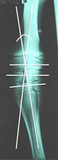
- Radiographs (Figure 5) to assess:
- The location of the deviation
- The degree of deviation from normal
- The condition of the bones within the affected joint
- Appearance of the growth plates
- Response to treatment over time
After assessment of your foal, your veterinarian may recommend one or more of the following treatment options:
1) Non-surgical treatments (considered for those foals that are very young and/or have a mild deviation). Non-surgical treatment options include:
- Stall rest
- Splints and/or cast placement on the affected leg
- Corrective hoof trimming and glue-on shoes with outside or inside extensions
2) Surgical treatments: (considered for those foals which have not responded to conservative management, those with severe deviations, and older foals whose affected bone has already completed its rapid growth phase). Surgical treatment options include:
- Periosteal Stripping: elevation of the periosteum of the bone to increase growth of one side of the affected bone (growth acceleration)
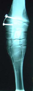
- Transphyseal Bridging: placement of screws & wires to slow growth of one side of the affected bone (growth retardation, Figure 6)
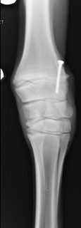
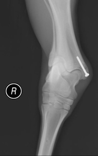
- Transphyseal Screw Placement (growth retardation, Figure 7)
- Corrective Osteotomy or Ostectomy: If the growth plates have closed and/or rotational or long bone deviations are present, a wedge of bone is removed and remaining bone is stabilized using bone plates and screws.
Restrictions and care following treatment depends on the procedure performed and severity of deviation, but may include:
- Stall rest +/- short amounts of controlled handwalking
- Daily monitoring of the affected limb(s) for correction in addition to heat, swelling, and lameness
- Maintaining a bandage over the surgical site until skin suture removal (10-14 days after surgery)
- Repeat radiographs to verify complete correction of the deviation
- Removal of screws and wires as soon as a visual correction of the deviation has occurred
Minor post-op complications can include:
- Incisional infection
- Seroma (an accumulation of fluid under the incision)
Major post-op complications can include:
- Overcorrection is possible with growth retardation (transphyseal bridging, transphyseal screw) procedures if the implants are not surgically removed at the appropriate time (or at all)
- Bone infection if the incision, screws, and/or wires become infected
The outcome of cases with angular limb deviation varies greatly, depending on the limb affected (front vs. hind), underlying condition, degree of deviation, joint affected, and expected future use of the horse.
Premier Sponsors


