The sinuses are air-filled cavities within the head of the horse. The sinuses also accommodate some of the maxillary premolar and molar tooth roots (upper cheek teeth), facilitate passage of facial nerves, and extend around (above and below) the horse’s eyes and end around the facial crest. The horse’s head has uniquely adapted itself and developed six pairs of paranasal sinuses—the frontal, sphenopalatine and maxillary sinuses, and the dorsal, middle and ventral conchal sinuses. The maxillary sinus is the largest paranasal sinus and is divided into two parts (rostral and caudal) by a thin septum. Many of the sinuses also communicate with one another: the maxillary sinus communicates with the frontal sinus, and the frontal sinus has a large communication with the dorsal conchal sinus. In healthy horses, mucus produced by the lining of the sinuses flows freely through the sinuses and into the nasal passages. The bone overlying the sinuses is very thin, and can be easily distorted by disease.
Sinusitis refers to inflammation or infection of one or more of the paranasal sinuses, and it is the most commonly encountered disease of the paranasal sinuses. It is classified as either primary or secondary, and acute or chronic. Primary sinusitis is defined as an infection in the sinus, usually bacterial in origin, which results in a buildup of pus within the sinus. Primary sinusitis is typically the result of an infection in the upper respiratory tract and it is most frequently caused by Streptococcus species of bacteria. It usually involves all paranasal sinus cavities, but may be confined to the ventral conchal sinus. More commonly however, is secondary sinusitis. Secondary sinusitis is an infection of the paranasal sinuses as a result of another primary cause, such as tooth root infection, bone fracture, or sinus cyst. The last four cheek teeth are most likely to cause a secondary sinusitis, as these teeth are contained within the maxillary sinuses. Empyema refers to purulent exudate within the sinus, but is not necessarily synonymous with chronic sinusitis.
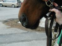
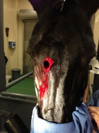
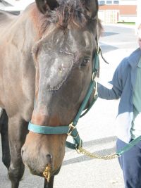
The most common sign of sinusitis (either primary or secondary) is nasal discharge. The nasal discharge usually occurs on the side of the affected sinus (unilateral) only (Figure 1). The appearance and character of the discharge is variable, and may contain pus or blood, with or without an odor. Other clinical signs include:
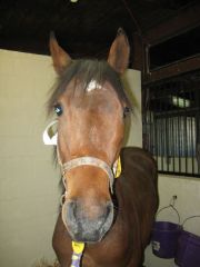
- Unilateral facial swelling (Figure 2)
- Epiphora (tearing of the eye
- Dull percussion of the sinuses
- Inspiratory noise
Your veterinarian will start by performing a complete physical examination before any other diagnostics. Additional diagnostics may include.
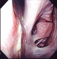
- Endoscopy: Endoscopy of the nasal passages and throat can help characterize where the nasal discharge is originating. Purulent material, a mass, or blood can sometimes be seen in the nasal passages originating from the nasomaxillary opening (Figure 3).
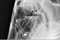
- Radiographs (X-rays): Head radiographs may reveal fluid lines, sinus cysts, solid masses, or changes in the surrounding bone associated with dental disease or neoplasia (Figure 4). If there is a large amount of exudate, it may be difficult to identify the cause of the sinusitis on radiographs, and the exudate may need to be removed to enhance radiographic evaluation.
- Sinocentesis: Sinocentesis involves making a small hole through the bone into the sinus and is used to obtain a sample of fluid from the sinus for examination and culture. This can usually be performed standing. The hole can also be used to lavage the sinus if purulent material (pus) is present.
- Sinoscopy: Sinoscopy is performed by making a small opening through the bone overlying the sinus and inserting a flexible endoscope into the sinus to evaluate (and potentially treat) the underlying condition. This is usually performed under standing sedation.
- Computed Tomography (CT): A CT scan can help characterize sinus disease most completely by providing a 3D image of the sinuses and surrounding structures. Typically CT scans require general anesthesia but they are rarely indicated in the initial stages of diagnosis.
Regardless of whether the sinusitis is primary or secondary, the goal of treatment is to treat the underlying cause of the sinusitis and to restore the horse’s natural sinus drainage mechanisms.
Primary paranasal sinusitis usually resolves with systemic antibiotic therapy and lavage. The exception is when the exudate becomes inspissated and obstructs appropriate flow through the nasal passages. In cases of secondary sinusitis, the primary disease must also be treated in order to fully resolve the sinusitis. Surgical treatments that may be used to remove exudate and provide additional drainage, if necessary, include sinus trephination and sinusotomy.
- Sinus trephination: Trephination of the affected sinus allows the sinus to be lavaged and antibiotics instilled directly into the sinus if indicated. Trephination is performed under standing sedation and local anesthesia. A small circle of bone overlying the affected sinus is removed (~10 mm) and a catheter can be inserted into the hole and maintained for multiple days to facilitate regular irrigation and flushing (Figure 5). Fluid and discharge produced from the nostrils is a good sign, and indicates a patent route for drainage from the sinus. The number of flushes depends on the degree and character of infection. Repeat endoscopy and lack of continued nasal discharge are two ways to assess the sinusitis has resolved..
- Sinusotomy: If the sinusitis persists or if good drainage cannot be established, more aggressive surgical management may be required. A sinusotomy, or bone flap is created under general anesthesia into the affected sinus (Figure 6). The sinus may be cleaned and appropriate drainage into the nasal passage can be established. Bleeding can be a complication of sinus surgery, but this depends on the exact cause of the sinusitis. The sinus may be packed to help reduce bleeding, and this pressure packing is typically removed a day or two after surgery through the nostril with the horse standing.
In cases of secondary sinusitis caused by dental disease, the affected tooth may be removed under standing sedation or general anesthesia. The tooth socket is typically flushed and packed with a non-absorbable material after removal to prevent recontamination of the sinus.
Anti-inflammatories are typically used for all cases of sinusitis, to help reduce any swelling or discomfort associated with the condition or surgical procedures. Ideally bacterial culture and sensitivity are used to select an appropriate antibiotic for lavage of the affected sinus(es). In some cases, systemic antibiotics or antifungal medications may also be indicated.
The duration of hospitalization for sinusitis depends on the underlying cause for the condition, and ranges from less than a day to several days. Repeat evaluation is likely needed to confirm the sinusitis has completely resolved. If the cause for the sinusitis was related to a tooth, repeat dental evaluation will likely be necessary.
Overall, the cosmetic results of equine sinus surgery are usually good. A small depression may be palpable at the surgical site, but is typically not noticeable to the naked eye. Horses with primary or secondary sinusitis typically have a good to excellent prognosis for return to function/athletic performance after appropriate treatment. If the cause of the sinusitis is treated appropriately and completely, it is unlikely the condition will recur in the future.
Premier Sponsors


