Skin tumors are common in horses of all breeds and ages. They tend to be locally invasive and slow to metastasize (spread to other areas of the body). The three most common skin tumors in horses are:
- Sarcoids
- Melanomas
- Squamous Cell Carcinoma
Many other types of tumors can also occur but are not as common. Early recognition, accurate diagnosis, and early treatment are crucial to obtaining a positive outcome after treatment. Delayed recognition and treatment increases the chances of recurrence or metastasis.
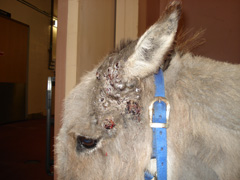
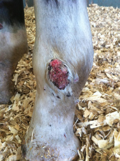
Sarcoids
Sarcoids are the most common type of skin tumor in horses which can occur in horses of any age. It is most common in adult Quarter Horses and other closely related breeds but rare in Standardbreds. There are four different forms of sarcoids; ranging from flat, ulcerated areas without hair (Figure 1), small nodules under the skin to large masses that can be haired, or look like proud flesh (Figure 2). They can occur on any part of the body, sometimes at areas of old wounds or repeated trauma such as corners of the cheek due to the bit and girth area.
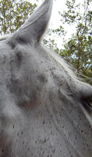
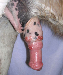
Melanomas
Melanomas occur almost exclusively in older grey horses. They are most commonly found under the tail, around the anus/perineum region, around the eyelids, in the parotid gland (at the throat latch region) and within the external/internal sheath in male horses. They can be black raised masses or raised masses underneath normal skin (Figure 3) and more likely to be malignant when found on non-grey horses.
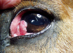
Squamous Cell Carcinomas
Squamous Cell Carcinomas are most commonly in adult to geriatric horses of any breed. They can be ulcerative or proliferative masses that look like proud flesh. They are common in un-pigmented (white or pink) areas, such as around the eye or third eyelid (Figure 4) and urogenital structures (Figure 5).
The best chance of treating skin tumors without recurrence or metastasis is through early diagnosis and treatment. Your primary care veterinarian may perform a physical examination and may perform a biopsy of the mass if large or complete excisional biopsy, if small. Your veterinarian should check for any of the following: lumps or masses on the skin, ocular discharge, bleeding from the penis, and difficulty urinating or defecating. Your veterinarian may choose to refer you to an ACVS board-certified veterinary surgeon for surgical removal depending on the location, temperament of the horse, the need for other therapies and whether or not specialized anesthetic techniques are required for complete removal of the tumor.
Treatment varies depending on the type of tumor, tumor location, and economic concerns. Most treatments consist of surgical removal +/- some form of additional therapy aimed at preventing re-occurrence or killing any residual tumor cells. Additional therapies include cryotherapy, laser, photodynamic therapy, radiation therapy, brachytherapy, and chemotherapy drugs ranging from topical creams to oral medications. Horses do not appear to suffer the negative side effects from radiation or chemotherapy that humans do and their quality of life during treatment is commonly excellent.
ACVS board-certified veterinary surgeons have expertise in complete surgical removal of skin tumors while obtaining the best cosmetic results possible. They also have access to the latest and most advanced treatments such as radiation therapy.
Aftercare will vary with the area involved and whether or not any additional therapy was required. If surgery was performed, then a short period of stall rest may be required to allow the incision to heal. The prognosis for most tumors is usually good if the tumor is treated early before it is too large. Sarcoids are very prone to recurring if they aren’t removed completely at the first surgery. Skin tumors are very slow to spread to other areas of the body (metastasize), but the risk of metastasis increases if they grow very large or are present for a long time.
Premier Sponsors


