The urinary tract includes the kidneys, ureters (small tubes that drain urine from the kidneys to the bladder), the bladder, and the urethra (the tube that carries urine from the bladder and out of the animal). Urolithiasis — the formation of “stones” or concretions of mucus, protein, and minerals in the urinary tract — is a common problem in male small ruminants and a frustrating one for owners and veterinarians.
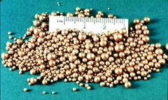
The composition of uroliths (also called “urinary stones” or “calculi”) varies with the part of the country that the animal lives in. The most common uroliths are calcium apatite and phosphatic-based calculi (for example, calcium hydrogen phosphate dihydrate, and magnesium ammonium phosphate or struvite). Silicate and calcium carbonate uroliths also are occasionally seen.
Urethral obstruction caused by uroliths (Figure 1) is most commonly seen in show or pet goats and lambs that have a high grain, low roughage diet. Diets that are high in grain, phosphorus, and magnesium and low in roughage (hay or fresh grass) and calcium will increase the risk of phosphate urolith formation. Normally, a ruminant will remove phosphorus from their body by excreting it into saliva and then out through the feces (manure). High grain, low roughage diets decrease the formation of saliva, so extra phosphorus must be removed from the blood by the kidneys and then excreted in the urine. When diets are too high in phosphorus, the urine phosphorus levels become excessively high, and the phosphorus settles and consolidates into stone-like pellets that can be too large to pass. These uroliths increase the risk of urinary tract infection and may result in life-threatening blockage of the urethra. Some breeds of sheep (for example, Texel and Scottish Blackface) may be predisposed to stone formation because they tend to excrete phosphorus through the urinary tract rather than the saliva and feces.
Early clinical signs include:
- Blood in the urine
- Straining to urinate
- Decreased urine production
- Painful urination
- Prolonged urination
- Dribbling urine
- Tail flagging
- Abdominal pain (stretching out all four limbs, kicking at the abdomen, looking at the side)
Late clinical signs include:
- Loss of appetite
- Lethargy (apparent depression)
- Abdominal swelling (from a ruptured bladder)
- Swelling around the prepuce (the skin covering the penis)
Once the animal is severely ill, they may lay on their side and not get up, and may eventually seizure or die suddenly.
Your veterinarian will perform a physical examination. Abnormal findings may include increased heart rate, rapid breathing, and possibly a large bladder. If the bladder has ruptured into the belly, a fluid wave can sometimes be felt (“balloted”) when pressure is put on one side of the abdomen.
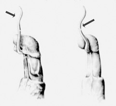
Most uroliths in small ruminants lodge at the “urethral process” or “vermiform appendage” — a small tube-like extension of skin and urethra at the tip of the penis (Figure 2); the second most common site is at the “distal sigmoid flexure” — an S-shaped curve in the lower half of the penis. Uroliths trapped in the urethral process can often be felt during physical examination.
As toxins build up, blood tests may show high concentrations of blood urea nitrogen (BUN), creatinine, and potassium, which are normally excreted in the urine. Other blood work changes may include high concentrations of muscle enzymes and low concentrations of sodium and chloride. Acid may also build up in the blood stream.
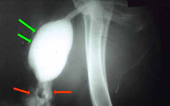
Uroliths are often diagnosed with ultrasound of the belly. The bladder and possibly the urethra can be distended (overfilled with fluid) on ultrasound, and sometimes the stones can be seen. Some uroliths are visible on radiographs (x-rays), which can be used to determine the number and location of stones. Special contrast studies can be performed with radiographs to determine if the bladder has ruptured (Figure 3).
Surgery is usually required to remove uroliths or relieve the blockage. Farm management practices should also be reviewed to determine whether diet or other factors could increase the risk of urolithiasis within the herd.
- Urethral Process Amputation:
The urethral process is a very short, narrow tube-like structure on the tip of the penis, and because of its location and size is the most common site for uroliths to obstruct. “Amputation” or removal of this process with a scalpel blade may allow uroliths within this process to pass, but recurrence of obstruction is likely, particularly if uroliths are also present higher up in the urethra. If this is the case, additional surgical procedures are needed. - Perineal Urethrostomy:
The urethra and its surrounding tissues can be easily felt in the back end (“perineum”) of sheep and goats 2-3 inches below the anus and just behind the rear legs. When uroliths block the penis or when the urethra has ruptured downstream from this area, a new opening can be made in this location to allow the animal to urinate like a female. This procedure is sometimes performed under heavy sedation with regional anesthesia (nerve blocks), but in valuable animals or pets it is often performed under general anesthesia. An incision is made through the skin and into the urethra, and the new urethral opening is sewn directly to the skin. The animal then urinates down and backwards instead of forwards. - Tube Cystostomy:
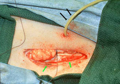
In animals with urethral obstruction or rupture, urine must be diverted away from the urethra to allow swelling to subside and tissues to heal. Tube cystostomy involves surgically placing a rubber tube in the bladder and exiting the tube through the belly wall adjacent to the prepuce (Figure 4). This procedure is performed in conjunction with urethral process amputation and surgical removal of uroliths from the bladder. Once the swelling in the urethra has resolved, any uroliths that were not removed at surgery often pass in the urine either through the urethra or the tube. Once the animal is healed, the tube can be removed. Although a tube cystostomy may be performed with sedation and local nerve blocks, general anesthesia is often used. This permits optimal sterile technique and time to remove uroliths from the bladder or repair it if it has ruptured and allows the surgeon to thoroughly flush the abdomen before placing the tube. Once the tube is in place, it can be attached to sterile collection system or covered with a one-way valve to allow urine excretion. - Prepubic Cystostomy:
For animals that have strictured (narrowed) perineal urethrostomy sites and subsequent re-obstruction, a permanent opening can be made between the bladder and the belly wall near the prepuce. This procedure is also called bladder “marsupialization”.
- Urethral Translocation:
This complicated procedure involves attaching the urethra in the belly to the lower half of the penis or prepuce. It can be attempted to “by-pass” a ruptured urethra (or failed urethrostomy).
-
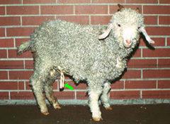
Figure 5: A ram with a cystostomy tube in place. The tube will be removed once the ram can urinate normally from the penis. The tube will be temporarily blocked before it is removed to make sure that the urethra is not blocked. Urethral Process Amputation: Usually minimal to no aftercare with excellent outcome if the blockage was only localized to the urethral process
- Perineal Urethrostomy: Daily evaluation of the site to ensure that the newly created hole remains open and has not strictured (closure of the hole) down in some animals. Also, the animal can no longer be used for breeding since semen does not pass through the penis.
- Tube cystostomy: The tube is kept in the animal until urine is seen dripping from the prepuce for 48 hours (Figure 5). Then, the tube is clamped and urination is monitored. If the animal can urinate, is not painful, and is able to empty its bladder, the tube is removed. If the animal can only partially urinate and continues to retain urine, the tube is left unobstructed for 5 to 7 more days and the process is repeated. On average, a tube cystostomy is removed 10 to 14 days after surgery. The main complication with a tube cystotomy is the risk of reobstruction following removal of the tube. Also, early removal (less than 5 days after placement) or accidental obstruction of the tube could result in leakage of urine into the abdomen (Figure 6).
-
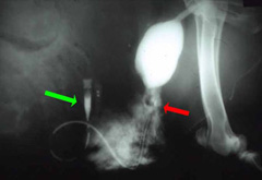
Figure 6: The cystostomy tube (green arrow) has accidentally been pulled out of the bladder and is now leaking urine (red arrow) into the abdomen. Prepubic Cystostomy: Urine will leak constantly from this hole, and may irritate the skin (“urine scald”), particularly if the skin around the new opening is not shaved and cleaned frequently. Development of chronic urinary tract infections. Therefore, this procedure is considered one of the “last resort” treatment for pet animals. In addition, the permanent cystostomy can become strictured and obstructed, requiring a second and sometimes a third surgical procedure.
- Urethral Translocation: This also is a “last resort” procedure for pet animals. Complications include loss of bladder tone and function (“neurogenic bladder atony”, with pooling of urine in the bladder and subsequent development of chronic cystitis (bladder infection).
Breeding males should be rested sexually for 1-2 weeks after urethral process amputations and for 2 months after other urethral surgeries to reduce the chance of surgical complications.
Recurrence is not uncommon, and animals must be monitored closely going forward. Prevention of urolithiasis involves ensuring ad lib water is available, feeding a high forage diet (avoiding high grain/concentrate diets), ensuring a balanced calcium-to-phosphate ratio in the diet, and adding salt (up to 3%) to promote drinking water. Urine acidifying diets or additives (ammonium chloride) can also be considered. Owners should always speak with their veterinarian and nutritionist prior to making changes to diet. It has also been suggested that delaying castration to a later age (12 weeks) may also protect development of the urethra and prevent occurrence of obstructive urolithiasis.
Photos provided courtesy of Susan L. Fubini, DVM, Diplomate ACVS and Peter C. Rakestraw, VMD, MA, Diplomate ACVS.
Premier Sponsors


