Colic in adult cattle can result due to derangements with any of the organ systems within the abdomen. Often colic is due to a problem within the gastrointestinal tract, however the peritoneum (tissue lining the abdomen), reproductive tract, and/or urinary tract can also be involved.
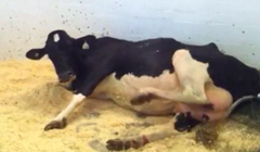
Colic in adult cattle can present with a variety of clinical signs, depending on the underlying cause (Figure 1). These signs can range from quite subtle to obvious. These signs can include:
- Acute drop in milk production
- Decreased feed intake to complete lack of feed intake
- Scant to no manure production
- Straining to defecate
- Abdominal distention
- Increased heart rate
- Increased respiratory rate
- Teeth grinding
Signs of abdominal discomfort:
- Kicking at abdomen
- Getting up and down multiple times
- Laying in lateral
- Flank watching
Obtaining an accurate history is key to appropriately guiding a diagnostic plan. Important information includes:
- Lactation number
- Days in milk
- Calving history
- Pregnancy status
- Diet
- Recent feed intake, appetite
- Manure production
- Previous medical history, including previous surgeries, and previous medications administered
A thorough physical examination should be performed. Simultaneous auscultation and percussion with particular attention paid to areas of resonance, abdominal palpation per rectum, and abdominal ballottement.
- Blood work including a packed cell volume, total protein, lactate, serum chemistry, and fibrinogen
- Urinalysis with particular attention paid to presence of ketones
- Additional diagnostics that can be performed to aid in obtaining an accurate diagnosis and/or surgical plan include:
- Imaging
- Abdominal radiography: most helpful if traumatic reticuloperitonitis is suspected
- Abdominal ultrasound: helpful when trying to determine presence of free peritoneal fluid, a specific organ or organ system involved, and for surgical planning/surgical approach
- Abdominocentesis: can be helpful, however cattle tend to compartmentalize peritonitis and fluid obtained may not be representative of the disease process
- Exploratory laparotomy: surgery can be used as a diagnostic tool to precisely characterize the cause of colic
- Imaging
Medical management can be used in the initial course of the disease
- Enteral fluids with laxatives (magnesium sulfate (MgSO4), mineral oil) and/or electrolytes
- Intravenous fluids
- Analgesics (flunixin meglumine/Banamine)
- Transfaunation
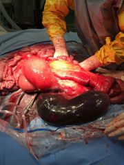
When medical management fails to resolve clinical signs, or if clinical signs indicate a surgical problem, surgical management is recommended. Surgical management includes an exploratory laparotomy where the abdomen and the organ systems are systematically examined. An exploratory laparotomy in a cow is most commonly performed through a flank incision and can be either done while standing with local anesthesia or under general anesthesia (Figure 2). Depending on the suspected cause of colic, an incision will be made in either the right or left flank in order to access specific areas within the abdomen. Conditions which cause colic in adult cattle and may require surgery include:
- Gastrointestinal:
- Traumatic reticuloperitonitis
- Abomasal displacement or volvulus
- Small intestinal obstructions
- Hemorrhagic bowel syndrome
- Small intestinal volvulus (mesenteric root, segmental)
- Intussception
- Foreign body (Trichobezoar, phytobezoar)
- Cecal dilatation or volvulus
- Spiral colon obstruction
- Peritonitis
- Reproductive:
- Uterine torsion
- Uterine tear
- Dystocia
- Urinary:
- Pyelonephritis
- Obstructive urolithiasis (typically in males)
- After exploratory laparotomy, hospitalization and continued treatments (intravenous fluids, antimicrobials therapy, and gradual return to feed) is often required for at least 3–5 days. Certain conditions and surgical procedures may require longer periods of hospitalization.
- Isolated box rest for at least 2 weeks is also required for appropriate healing of the laparotomy incision.
- Monitoring attitude, appetite, and manure production are important once the animal leaves the hospital. Monitoring the incision for increased heat, swelling, and discharge should also be performed daily.
- Depending on the condition of the cow at the time of presentation and depending on the cause of the colic, outcome and prognosis can be quite variable.
- Surgical correction of displaced abomasums have a fair-good prognosis:
- 86–90% success rate of left displaced abomasums corrected surgically with omentopexy
- 68–74% success rate of right abomasal volvulus corrected surgically
- Hemorrhagic bowel syndrome has a poor prognosis with a high rate of recurrence
- Dennison et al, 2002
- Medical management- 1/8 (13%) survived
- Surgical management- 3/13 (23%) survived
- Dennison et al, 2002
- Surgical correction of displaced abomasums have a fair-good prognosis:
- Cecal dilatation/volvulus generally has a good prognosis if the condition is treated early, prior to any compromise of the cecum. Braun et al, 2012: 87% responded to conservative or surgical management.
Premier Sponsors


