Congenital ear canal atresia (CECA) is a birth defect that results in failure of normal formation of the outer ear canal opening and is a rare diagnosis in dogs. Early in life, this finding may go undiagnosed and does not initially pose major risks. However, as the ear canal is incomplete, the normal wax and skin cells that expel from the ear opening become entrapped, this defect can result in a swelling of the ear canal, infection, abscessation, hearing loss, and neurologic changes. Early identification of this defect by having your veterinarian perform a complete physical examination and pursuing surgical intervention has been shown to reduce the development of severe changes and result in preserved hearing.
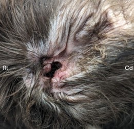
At birth, all dog ears appear closed, and an external ear canal opening is not visible. As dogs grow, the external ear canal opens. This occurs around the age of two weeks. Failure of the ear canal to open may indicate that your pet has CECA. Your pet will likely have their first veterinary visit around four weeks of age and a good physical examination is imperative to evaluate for this defect
(Figure 1) along with potential other congenital anomalies. Your pet may not be showing obvious signs related to hearing or ear discomfort as these symptoms develop with chronicity. If CECA is identified and not treated, your dog will likely develop clinical signs of pain or discomfort including trying to scratch at their ear, shaking their head, and potentially developing a head tilt.
It is important to avoid delaying intervention to keep your pet pain free and avoid complications such as infection or abscess formation, hearing loss, and neurologic changes. Consultation by an ACVS board-certified veterinary surgeon is recommended to evaluate your pet for CECA.
You may be able to identify that your pet is lacking an external ear canal opening at home. If you believe that your pet might have CECA, then additional diagnostics are indicated. The first step is imaging to assess what the entire ear canal looks like. A CT scan is helpful in determining how the defect has affected the surrounding anatomy and help guide the veterinary surgeon in deciding which surgical intervention is most appropriate. Unfortunately, it is impossible to determine by imaging alone if symptoms such as hearing loss will be permanent. Bloodwork to evaluate the overall health of your pet prior to undergoing anesthesia will likely be recommended.
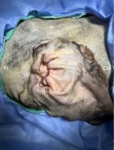
Based on your veterinary surgeon’s evaluation of your pet’s clinical status and CT findings, surgical options will be discussed. A
reconstructive surgery to create a new external ear canal opening (Figure 2) may be possible and can result in preservation of hearing. If reconstructive surgery is indicated, this can be followed by video imaging of the ear drum and ear canals by a board-certified veterinary dermatologist. This collaboration can help to ensure that, following re-establishment of the ear canal, any potential infection is appropriately treated.
For patients with long-standing CECA and potential chronic irreversible changes, a TECA-LBO, surgical full removal of the ear canal resulting in loss of hearing may be recommended.
For two weeks following surgery, close incision monitoring and activity restriction is critical to prevent infections. Once the incision is healed, follow-up with a veterinary dermatologist may be indicated. The risk of long-term neurologic signs is more common following TECA-LBO intervention compared to reconstructive surgery and may require therapies such as regular eye lubrication administration. Overall, dogs respond well to these surgical interventions and underlying pain resolves making your pet more comfortable to resume normal activities.
Premier Sponsors


