A cleft palate is an opening between the mouth and the nose that happens when the tissues separating these two cavities do not grow together properly. This birth defect can occur in the lip (primary cleft palate, cleft lip, or harelip) or along the roof of the mouth (secondary cleft palate). Within the mouth, the cleft, or opening, can extend along the bony portion (hard palate), the flexible portion used in swallowing (soft palate), or both.
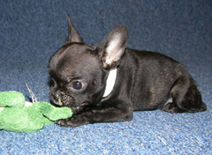
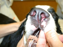
Purebred dogs and cats have a higher incidence of cleft palate, and brachycephalic breeds, with their short stubby faces, are most commonly affected (Figure 1). Cleft palates may occur more commonly in Boston terriers, Pekingese, bulldogs, miniature schnauzers, beagles, cocker spaniels, dachshunds, and Siamese cats. Although genetics is considered the main cause of this problem, nutritional deficiencies, viruses, and poisons that affect the mother during pregnancy may also increase the risk of cleft palates.
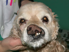
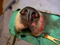
Pets with primary cleft palates are obvious (Figure2).
- Teeth and gums of the upper jaw may be showing.
- One incorrectly shaped nostril.
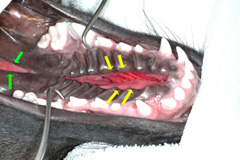
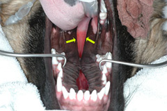
Pets with secondary cleft palates (Figure 3) within the mouth may:
- Sneeze and snort because food and saliva will pass into the nose
- Have a “runny” nose after eating, or before or after nursing. It will become a constant drip if it becomes infected.
- Cough and gag when they drink water
- Not grow well due to trouble eating
- Have trouble breathing and exercising because of fluid or infection in their noses.
- Oral examination: Cleft palate of the lip and the hard palate are easy to see. Most pets are anesthetized; however, to see the soft palate because it is so far back in the mouth.
- Chest x-rays to look for signs of pneumonia.
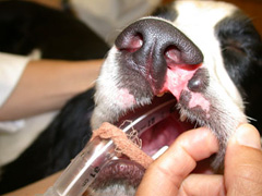
Small primary clefts of the lip and nostril rarely cause clinical problems, but they are unsightly and most pet owners prefer to have those corrected (Figures 4, 5, 6).
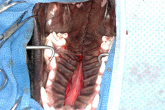
Secondary cleft palates require surgical treatment to prevent long-term nasal and lung infections and to help your pet receive proper nutrition. (Figures 7, 8)
The surgery is difficult on very young pets and, with growth, the cleft in hard palates may become smaller, so puppies and kittens are often fed with feeding tubes until they reach 3-4 months of age. Pet owners can learn how to pass a feeding tube at each meal or their primary care veterinarian can place a feeding tube through the side of the neck to easily feed blenderized diets.
Because the surgery is performed in young, underweight pets with breathing problems, anesthesia and surgery recovery can be risky. Many pets have swelling of the soft palate after the surgery, which can cause breathing problems or snoring, is usually resolve.
Restrictions following surgery usually are:
- Give antibiotics to pets with pneumonia or nasal infections.
- Leave e-collars on for 1-2 weeks to stop pets from rubbing their faces.
- Feed soft, blenderized foods by mouth or through a feeding tube for 2–4 weeks after surgery.
- Do not give hard food or toys for at least a month.
Postoperative complications can include:
- Partial surgical site failure (called dehiscence), or opening due to tension or the puppy/kitting chewing or pawing at their face
- Nasal discharge or sneezing
- Continued coughing and gagging due to a short soft palate
Prognosis is excellent for pets with small clefts. When more than half of the hard palate is affected, surgery is much more difficult and more complications are expected. Very large defects are closed with special dental appliances and tissue techniques.
Do not bred pets that are born with cleft palates, and their parents should not be bred if they are one of the predisposed breeds.
Refer pets with secondary cleft palates to an ACVS board-certified veterinary surgeon for repair. The greatest success rates are seen when the procedure is performed initially by an experienced veterinary surgeon.
Premier Sponsors


