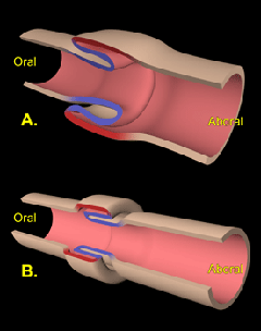
Intussusception (pronounced in-tuh-sus-sep-shun) describes a condition in which one segment of the intestine (the intussusceptum) telescopes or invaginates (Figure 1) into the lumen of an adjacent segment of intestine (the intussuscipiens).
Intussusceptions may occur at any location in the gastrointestinal tract from the stomach to the large intestine. However, most commonly, the bowel segments involved are the middle of the small intestine (jejunum ) or the where the small intestine joins the large intestine or colon (ileocecocolic junction). Generally the intussusceptum is a more proximal portion of bowel (i.e., closer to the mouth) which telescopes into a more distal (i.e., closer to the anus) segment. This pattern follows the normal direction of peristalsis. The reverse, however, is occasionally found.
Intussusceptions are most commonly associated with some problem that causes inflammation of the intestine (enteritis). Common causes of enteritis are:
- intestinal parasites (hookworms, whipworms, and roundworms)
- protozoal, bacterial or viral infections (Giardia, Salmonella, canine distemper, and parvovirus)
- intestinal foreign bodies (bones, plastic toys, etc.)
- abrupt dietary changes
- intestinal masses (tumors)
- a surgical procedure performed on the intestine
Increased motility in a segment of intestine (hypermotility) which is adjacent to a segment that has lack of motility (ileus) can cause the hypermotile segment to telescope into the segment with ileus, resulting in an intussusception.
Dogs and cats that develop intussusceptions have generally been having episodes of diarrhea or vomiting before the intussusception occurs. Small volumes of bloody diarrhea, abdominal pain, or a palpable abdominal mass are suggestive of an intussusception. The severity of the clinical signs depends somewhat on the location of the intussusception, with problems lower in the intestinal tract causing less severe clinical signs. Intussusceptions can be chronic or intermittent, meaning that they will reduce themselves spontaneously and then reform.
Dogs or cats with a history of vomiting or diarrhea for more than a day or two should be evaluated by your primary care veterinarian, particularly if associated with depression and loss of appetite.
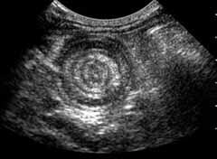
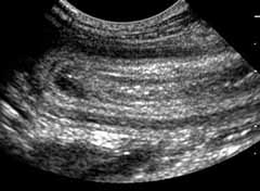
Intussusception should be a consideration if your pet has a history of vomiting or diarrhea that has a palpable mass in the abdomen. The mass can be felt as a thickened sausage-shaped intestinal loop. Occasionally the small bowel can be felt entering the mass. Radiographs will show a typical pattern of intestinal obstruction with gas and fluid-filled dilated loops of bowel if the obstruction caused by the intussusception is complete. In cases of partial obstruction, there may not be significant signs on plain radiographs and a barium contrast study may be needed to identify the problem. Ultrasound examination of the abdomen is very useful for identifying the intussusception area (Figures 2a and 2b).
Because most pets that develop intussusceptions have had episodes of vomiting and diarrhea, the hydration and electrolyte status should be addressed prior to surgery if possible. This involves some blood chemistry analysis and treatment with an appropriate intravenous fluid. Treatment of animals with intussusception can be complicated and difficult. Many veterinarians prefer to send these patients to an ACVS board-certified veterinary surgeon for ongoing care.
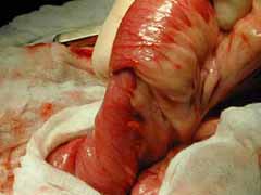
Occasionally intussusceptions can be manually reduced by manipulation of the affected bowel through the abdomen or they reduce themselves spontaneously. In most cases, however, surgery is required to treat this problem. Recurrence of intussusceptions is common, so even if the intussusception can be manually reduced, surgery is often recommended to perform procedures designed to decrease the incidence of recurrence. During surgery the affected area bowel is easily identified (Figure 3). It is occasionally possible for the surgeon to manually reduce the intussusception. In many instances, either the intussusception cannot be reduced or the bowel is so badly damaged that resection of the affected bowel is required. In this case, the area of damaged bowel is removed and the cut ends of the intestine are joined together with sutures or staples in a procedure called an intestinal anastomosis.
Postoperative care following intussusception involves efforts to manage pain, generally with opioids, which help to slow bowel motility as well. Re-establishment of hydration and normal electrolyte values is essential and appropriate intravenous fluids are generally used until your pet is eating normally. Antibiotics may be required depending on the amount of contamination from the surgery and the preference of the surgeon.
The prognosis following surgical repair of an intussusception depends on several factors including:
- the duration of the problem
- the amount of intestine involved
- the location of the problem and
- the extent of the blockage that has been caused
Chronic intussusceptions usually require removal of a section of bowel and anastomosis of the ends to re-establish bowel integrity. Anytime bowel has to be removed there is a chance of leakage from the surgery site which can result in potentially fatal peritonitis. Pets that are in poor condition because of the intussusception may have a diminished ability to heal, making leakage more likely. If large amounts of bowel have to be removed, your pet may not do well because of the relatively short length of bowel left behind.
The prognosis for pets with an intussusception is good as long as recurrence of the problem can be prevented and excessive amounts of bowel do not have to be removed. It has been reported that between 11% and 20% of dogs will have a recurrence of the problem following surgical correction. The incidence is higher (25%) if only manual reduction and no surgery is done. A procedure known as enteroplication can be performed to prevent recurrence of the intussusception, however, may make the patient more susceptible to other complications such as intestinal obstructions with foreign material that may have been able to pass without complication if the bowel had not been plicated.
Premier Sponsors